

The Importance of Dermatoscopy for the Diagnosis of Melanonychia
- PMID: 31297260
- PMCID: PMC6592664
- DOI: 10.12865/CHSJ.45.01.04
The Importance of Dermatoscopy for the Diagnosis of Melanonychia
Abstract
Melanonychia is the brown or black color of the finger or toe nail due to melanin deposition or melanocytes in the nail plate. The evidence of melanocytic disease is made by the dermatoscope, which allows to highlight the anomalies of the plate. The purpose of our study was to evaluate dermatoscopically the melanonychia, both in the form of stain and longitudinal on finger and/or toe nails in order to establish the type of nail hyperpigmentation.
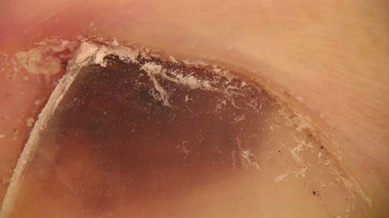
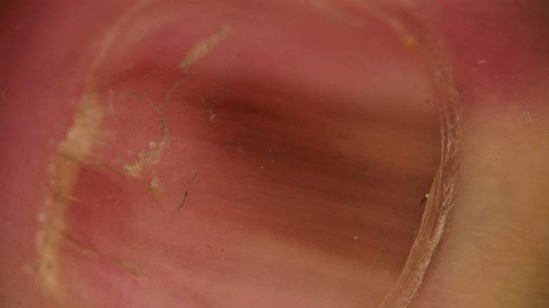
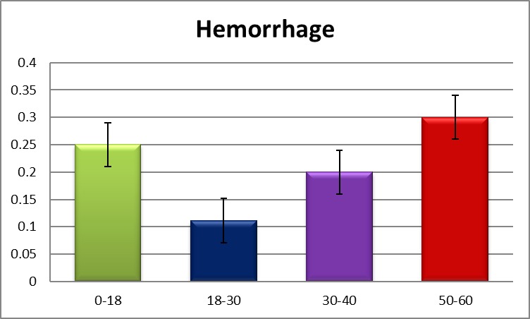
Materials and method: 33 patients with longitudinal and stain melanonychia were examined with 30x Molemax HD computerized dermatoscope between May 2017-septembre 2018 in this prospective study conducted in the Department of Dermatology of Medical Center Dr. Ianosi (Craiova, Romania). Clinical data included: type of melanonychia, number and name of involved fingers, the presence or absence of fungal infections, nail apparatus tumors or hemorrhage.
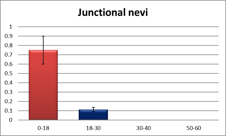
Results: The most frequent nail diagnosis was fungal infection (onychomycosis) observed in 18 patients (54.54%), malignant melanoma was diagnosed in 1 patient (3.03%) and the junctional nevus in 4 patients (12.12%). In 18 patients which has longitudinal melanonychia, the most frequent involved finger was the big toe, and in 15 patients which has stain melanonychia, all of them (100%) had affected the big toe, 7 (46.66%) patients had affected the thumb and the same percent the forth finger.
Conclusion: Nail dermatoscopy is an important method in establishing the diagnosis of melanonychia and allowed to avoid unnecessary biopsy for melanonychia.
Keywords: dermatoscopy; melanoma; melanonychia; onychomycosis.
Figures

References
-
- Argenziano G, Albertini G, Castagnetti F, De Pace, Di Lernia, Longo C, Pellacani G, Piana S, Ricci C, Zalaudek I. Early diagnosis of melanoma: What is the impact of dermoscopy? DermatolTher. 2012;25(5):403–409. - PubMed
-
- Astur M. M., Farkas C.B., Junqueira J.P., Enokihara M.M., Enokihara M.Y., Michalany N. Reassessing melanonychiastriata in phototypes IV, V, and VI patients. DermatolSurg. 2016;42(2):183–190. - PubMed
-
- Koga H. Yoshikawa S., Shinohara T., Le Gal F.A., Cortés B., Saida T. Long-term follow-up oflongitudinal melanonychia in children and adolescents using an objective discrimination index. ActaDermVenereol. 2016;96(5):716–717. - PubMed
-
- Koga H, Saida T, Uhara H. Key point in dermoscopic differentiation between early nail apparatus melanoma and benign longitudinal melanonychia. J Dermatol. 2011;38(1):45–52. - PubMed
Publication types
LinkOut - more resources
Full Text Sources