

ANKRD44 Gene Silencing: A Putative Role in Trastuzumab Resistance in Her2-Like Breast Cancer
- PMID: 31297336
- PMCID: PMC6607964
- DOI: 10.3389/fonc.2019.00547
ANKRD44 Gene Silencing: A Putative Role in Trastuzumab Resistance in Her2-Like Breast Cancer
Erratum in
-
Erratum: ANKRD44 Gene Silencing: A Putative Role in Trastuzumab Resistance in Her2-Like Breast Cancer.Front Oncol. 2019 Jul 18;9:709. doi: 10.3389/fonc.2019.00709. eCollection 2019. Front Oncol. 2019. PMID: 31380291 Free PMC article.
Abstract
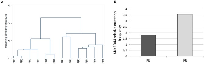
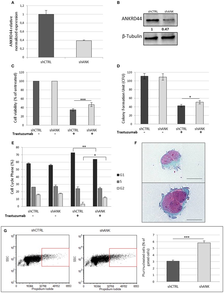
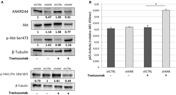
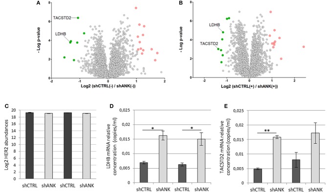
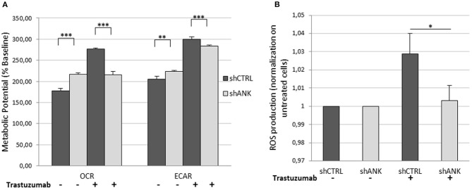
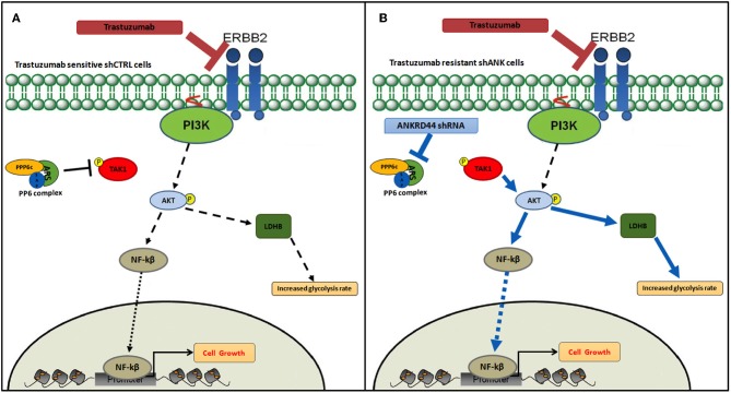
Trastuzumab is an effective therapeutic treatment for Her2-like breast cancer; despite this most of these tumors develop resistance to therapy due to specific gene mutations or alterations in gene expression. Understanding the mechanisms of resistance to Trastuzumab could be a useful tool in order to identify combinations of drugs that elude resistance and allow a better response for the treated patients. Twelve primary biopsies of Her2+/hormone receptor negative (ER-/PgR-) breast cancer patients were selected based on the specific response to neoadjuvant therapy with Trastuzumab and their whole exome was sequenced leading to the identification of 18 informative gene mutations that discriminate patients selectively based on response to treatment. Among these genes, we focused on the study of the ANKRD44 gene to understand its role in the mechanism of resistance to Trastuzumab. The ANKRD44 gene was silenced in Her2-like breast cancer cell line (BT474), obtaining a partially Trastuzumab-resistant breast cancer cell line that constitutively activates the NF-kb protein via the TAK1/AKT pathway. Following this activation an increase in the level of glycolysis in resistant cells is promoted, also confirmed by the up-regulation of the LDHB protein and by an increased TROP2 protein expression, found generally associated with aggressive tumors. These results allow us to consider the ANKRD44 gene as a potential gene involved in Trastuzumab resistance.
Keywords: ANKRD44; Her2+ breast cancer; LC-MS/MS; Trastuzumab resistance; gene silencing; next generation sequencing.
Figures
Similar articles
-
Erratum: ANKRD44 Gene Silencing: A Putative Role in Trastuzumab Resistance in Her2-Like Breast Cancer.Front Oncol. 2019 Jul 18;9:709. doi: 10.3389/fonc.2019.00709. eCollection 2019. Front Oncol. 2019. PMID: 31380291 Free PMC article.
-
YAP increases response to Trastuzumab in HER2-positive Breast Cancer by enhancing P73-induced apoptosis.J Cancer. 2020 Sep 25;11(22):6748-6759. doi: 10.7150/jca.48535. eCollection 2020. J Cancer. 2020. PMID: 33046997 Free PMC article.
-
Cullin7 enhances resistance to trastuzumab therapy in Her2 positive breast cancer via degrading IRS-1 and downregulating IGFBP-3 to activate the PI3K/AKT pathway.Cancer Lett. 2019 Nov 1;464:25-36. doi: 10.1016/j.canlet.2019.08.008. Epub 2019 Aug 25. Cancer Lett. 2019. PMID: 31461670
-
Evolving strategies for overcoming resistance to HER2-directed therapy: targeting the PI3K/Akt/mTOR pathway.Clin Breast Cancer. 2010 Nov;10 Suppl 3:S72-8. doi: 10.3816/CBC.2010.s.015. Clin Breast Cancer. 2010. PMID: 21115425 Review.
-
Recent Insights into the Development of Preclinical Trastuzumab- Resistant HER2+ Breast Cancer Models.Curr Med Chem. 2018;25(17):1976-1998. doi: 10.2174/0929867323666161216144659. Curr Med Chem. 2018. PMID: 27993109 Review.
Cited by
-
Recent Advances in Cancer Plasticity: Cellular Mechanisms, Surveillance Strategies, and Therapeutic Optimization.Front Oncol. 2020 Apr 22;10:569. doi: 10.3389/fonc.2020.00569. eCollection 2020. Front Oncol. 2020. PMID: 32391266 Free PMC article. Review.
-
Chemoresistance in breast cancer: PI3K/Akt pathway inhibitors vs the current chemotherapy.Am J Cancer Res. 2021 Oct 15;11(10):5155-5183. eCollection 2021. Am J Cancer Res. 2021. PMID: 34765318 Free PMC article. Review.
-
Targeting Glycolysis for Treatment of Breast Cancer Resistance: Current Progress and Future Prospects.Int J Biol Sci. 2025 Mar 24;21(6):2589-2605. doi: 10.7150/ijbs.109803. eCollection 2025. Int J Biol Sci. 2025. PMID: 40303296 Free PMC article. Review.
-
Circ-RNF111 contributes to paclitaxel resistance in breast cancer by elevating E2F3 expression via miR-140-5p.Thorac Cancer. 2020 Jul;11(7):1891-1903. doi: 10.1111/1759-7714.13475. Epub 2020 May 23. Thorac Cancer. 2020. PMID: 32445273 Free PMC article.
-
A Genetic Screen Identifies Etl4-Deficiency Capable of Stabilizing the Haploidy in Embryonic Stem Cells.Stem Cell Reports. 2021 Jan 12;16(1):29-38. doi: 10.1016/j.stemcr.2020.11.016. Stem Cell Reports. 2021. PMID: 33440180 Free PMC article.
References
-
- Hellstrom I, Goodman G, Pullman J, Yang Y, Hellstrom KE. Overexpression of HER-2 in ovarian carcinomas. Cancer Res. (2001) 61:2420–3. - PubMed
LinkOut - more resources
Full Text Sources
Research Materials
Miscellaneous