. 2019 Jun 26:9:231.
doi: 10.3389/fcimb.2019.00231. eCollection 2019.

Depletion of Host Cell Focal Adhesion Kinase Increases the Susceptibility to Invasion by Trypanosoma cruzi Metacyclic Forms

Affiliations

Depletion of Host Cell Focal Adhesion Kinase Increases the Susceptibility to Invasion by Trypanosoma cruzi Metacyclic Forms

Thiago Souza Onofre et al. Front Cell Infect Microbiol. .

Abstract

Focal adhesion kinase (FAK), a cytoplasmic protein tyrosine kinase (PTK), is implicated in diverse cellular processes, including the regulation of F-actin dynamics. Host cell F-actin rearrangement is critical for invasion of Trypanosoma cruzi, the protozoan parasite that causes Chagas disease. It is unknown whether FAK is involved in the internalization process of metacyclic trypomastigote (MT), the parasite form that is important for vectorial transmission. MT can enter the mammalian host through the ocular mucosa, lesion in the skin, or by the oral route. Oral infection by MT is currently a mode of transmission responsible for outbreaks of acute Chagas disease. Here we addressed the question by generating HeLa cell lines deficient in FAK. Host cell invasion assays showed that, as compared to control wild type (WT) cells, FAK-deficient cells were significantly more susceptible to parasite invasion. Lysosome spreading and a disarranged actin cytoskeleton, two features associated with susceptibility to MT invasion, were detected in FAK-deficient cells, as opposed to WT cells that exhibited a more organized F-actin arrangement, and lysosomes concentrated in the perinuclear area. As compared to WT cells, the capacity of FAK-deficient cells to bind a recombinant protein based on gp82, the MT surface molecule that mediates invasion, was higher. On the other hand, when treated with FAK-specific inhibitor PF573228, WT cells exhibited a dense meshwork of actin filaments, lysosome accumulation around the nucleus, and had increased resistance to MT invasion. In cells treated with PF573228, the phosphorylation levels of FAK were reduced and, as a consequence of FAK inactivation, diminished phosphorylation of extracellular signal-regulated protein kinases (ERK1/2) was observed. Fibronectin, known to impair MT invasion, induced the formation of thick bundles of F-actin and ERK1/2 dephosphorylation.

Keywords: Trypanosoma cruzi; actin cytoskeleton; focal adhesion kinase; host cell invasion; lysosome distribution; metacyclic trypomastigote.


Figures
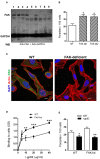
Figure 1
Figure 1
FAK knockdown increases host cell susceptibility to T. cruzi MT invasion and gp82 binding capacity. (A) HeLa cells were subjected to lentiviral transduction for FAK knockdown (kd) and then analyzed by western blotting (WB) using the indicated antibodies. Shown are the wild type (WT) cells (lane 1), cells transduced with lentivirus containing unrelated sequence (lanes 2 and 3), cells targeted with FAK sequence 1 (lanes 4, 5, 6) or with FAK sequence 2 (lanes, 7, 8, 9). Note the FAK depletion in four cell lines (lanes 5, 7, 8, 9). (B) WT cells and two FAK-depleted cell lines (FAK-kd1 and FAK-kd2) were incubated with MT for 1 h and then processed for intracellular parasite counting. Values are the means ± SD of three independent assays performed in duplicate. MT invasion was significantly increased in FAK-deficient cells (*P < 0.005). (C) WT and FAK-deficient cells were processed for immunofluorescence analysis using anti-FAK antibody, Alexa Fluor 488-conjugated anti-mouse IgG (green), TRITC-phalloidin (red) for actin detection and DAPI (blue) for DNA, under 63x objective. Scale bar = 10 μm. (D) WT and FAK-deficient cells, grown in microtiter plates, were fixed and incubated sequentially with r-gp82, anti-gp82 antiserum, and anti-mouse IgG conjugated to peroxidase. The binding of r-gp82, revealed with o-phenylenediamine and expressed as optical density value, was significantly higher in FAK-deficient cells than in WT cells (*P < 0.05, **P < 0.01, ***P < 0.001). A representative result of three independent assays performed in triplicate is shown. (E) WT and FAK-deficient cells were incubated with MT in the presence of r-gp82 or GST. Values are the means ± SD of three independent assays performed in duplicate. MT invasion was significantly decreased in the presence of r-gp82 (*P < 0.05, **P < 0.005).
Figure 2
Figure 2
FAK-deficient and WT cells exhibit distinct F-actin organization and lysosome distribution. WT and FAK-deficient cells were analyzed by confocal fluorescence microscopy, to visualize actin cytoskeleton (red), lysosomes (green), and nucleus stained with DAPI (blue), under 63x objective. Scale bar = 20 μm. Note the F-actin disarrangement and lysosome spreading in FAK-deficient cells.
Figure 3
Figure 3
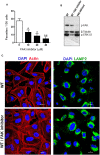
FAK inhibition in WT cells induces FAK and ERK1/2 dephosphorylation, and reduces MT invasion by affecting the actin cytoskeleton architecture. (A) Cells were untreated or treated for 45 min with FAK inhibitor PF573228 at the indicated concentrations, the drug was removed, parasites were added, and incubation proceeded for 1 h before intracellular MT counting. Values are the means ± SD of three independent assays performed in duplicate. MT invasion was significantly reduced in cells treated with FAK inhibitor (*P < 0.005, **P < 0.001). (B) Detergent-soluble extracts of untreated and FAK inhibitor-treated WT cells, as well as of FAK-deficient cells, were analyzed by western blotting, using antibody to phospho-FAK (Tyr397), phospho-ERK1/2 and β-tubulin. Note the decreased phosphorylation levels of FAK and ERK1/2 in FAK inhibitor-treated WT cells. (C) WT cells, treated or not with FAK inhibitor PF573228 and then processed for confocal fluorescence microscopy to visualize actin cytoskeleton (red), lysosomes (green), and nucleus (blue). Scale bar = 20 μm. Note the altered F-actin arrangement in FAK inhibitor-treated cells.
Figure 4
Figure 4
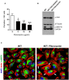
Fibronectin-induced decrease in MT invasion is associated with ERK1/2 dephosphorylation and F-actin bundle formation. (A) WT cells were pretreated for 30 min with fibronectin at the indicated concentrations, parasites were then added and the incubation proceeded for 1 h. After fixation and Giemsa staining, the internalized parasites were quantified in at least 250 cells/duplicate. Values are the means ± SD of four independent assays. MT invasion was significantly reduced in cells treated with fibronectin at all concentrations (*P < 0.01, **P < 0.005, ***P < 0.001). (B) Detergent-soluble extracts of cells untreated or treated with FAK inhibitor or fibronectin were analyzed by western blotting, using antibody to phospho-FAK (Tyr397), phospho-ERK1/2. Specific antibodies were used to reveal the loading controls β-tubulin and GAPDH. Note the decreased phosphorylation levels of ERK1/2, but not of FAK, in fibronectin-treated WT cells. (C) WT cells, treated or not with fibronectin, were processed for confocal microscopy to visualize actin cytoskeleton (red), lysosomes (green), and nucleus (blue). Scale bar = 20 μm. Note the dense F-actin bundles in fibronectin-treated cells (white arrows).
Figure 5
Figure 5
EA internalization is not significantly affected by FAK depletion or by treatment of WT cells with FAK inhibitor. (A) WT and FAK-deficient cells were incubated for 1 h with EA. (B) WT cells were treated or not with FAK inhibitor before incubation with EA. Intracellular parasites were quantified in at least 250 cells/duplicate. Values are the means ± SD of three independent assays. No significant difference was detected between FAK-deficient or FAK inhibitor-treated cells and the respective controls.

Similar articles

Cited by

References

    1. Agerer F., Lux S., Michel A., Rohde M., Ohlsen K., Hauck C. R. (2005). Cellular invasion by Staphylococcus aureus reveals a functional link between focal adhesion kinase and cortactin in integrin-mediated internalisation. J. Cell Sci. 118, 2189–2000. 10.1242/jcs.02328 - DOI - PubMed
    1. Bonfim-Melo A., Ferreira E. R., Florentino P. T. V., Mortara R. A. (2018). Amastigote synapse: the tricks of Trypanosoma cruzi extracellular amastigotes. Front. Microbiol. 9:1341. 10.3389/fmicb.2018.01341 - DOI - PMC - PubMed
    1. Bonfim-Melo A., Zanetti B. F., Ferreira E. R., Vandoninck S., Han S. W., Van Lint J., et al. . (2015). Trypanosoma cruzi extracellular amastigotes trigger the protein kinase D1-cortactin-actin pathway during cell invasion. Cell. Microbiol. 17, 1797–1810. 10.1111/cmi.12472 - DOI - PubMed
    1. Brener Z., Chiari E. (1963). Variacões morfol6gicas observadas em diferentes amostras de Trypanosoma cruzi. Rev. Inst. Med. Trop. Sao Paulo 5, 220–224. - PubMed
    1. Cortez C., Real F., Yoshida N. (2016). Lysosome biogenesis/scattering increases host cell susceptibility to invasion by Trypanosoma cruzi metacyclic forms and resistance to tissue culture trypomastigotes. Cell. Microbiol. 18, 748–760. 10.1111/cmi.12548 - DOI - PMC - PubMed

Publication types

MeSH terms

Substances