. 2019 Jul 11;21(5):89.
doi: 10.1208/s12248-019-0354-6.

Quality Controls in Ligand Binding Assays: Recommendations and Best Practices for Preparation, Qualification, Maintenance of Lot to Lot Consistency, and Prevention of Assay Drift

Affiliations

Quality Controls in Ligand Binding Assays: Recommendations and Best Practices for Preparation, Qualification, Maintenance of Lot to Lot Consistency, and Prevention of Assay Drift

Mitra Azadeh et al. AAPS J. .

Abstract

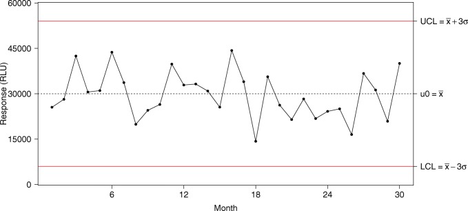
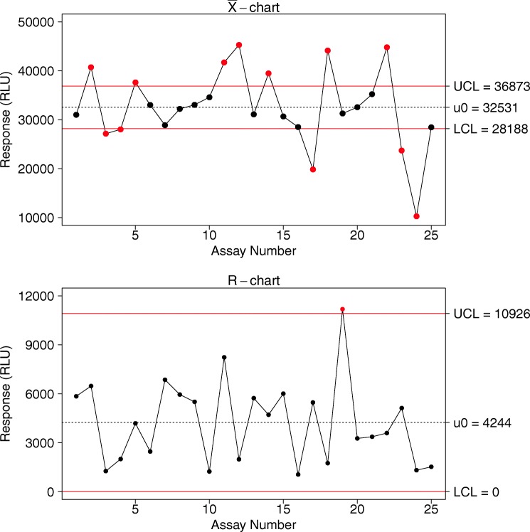
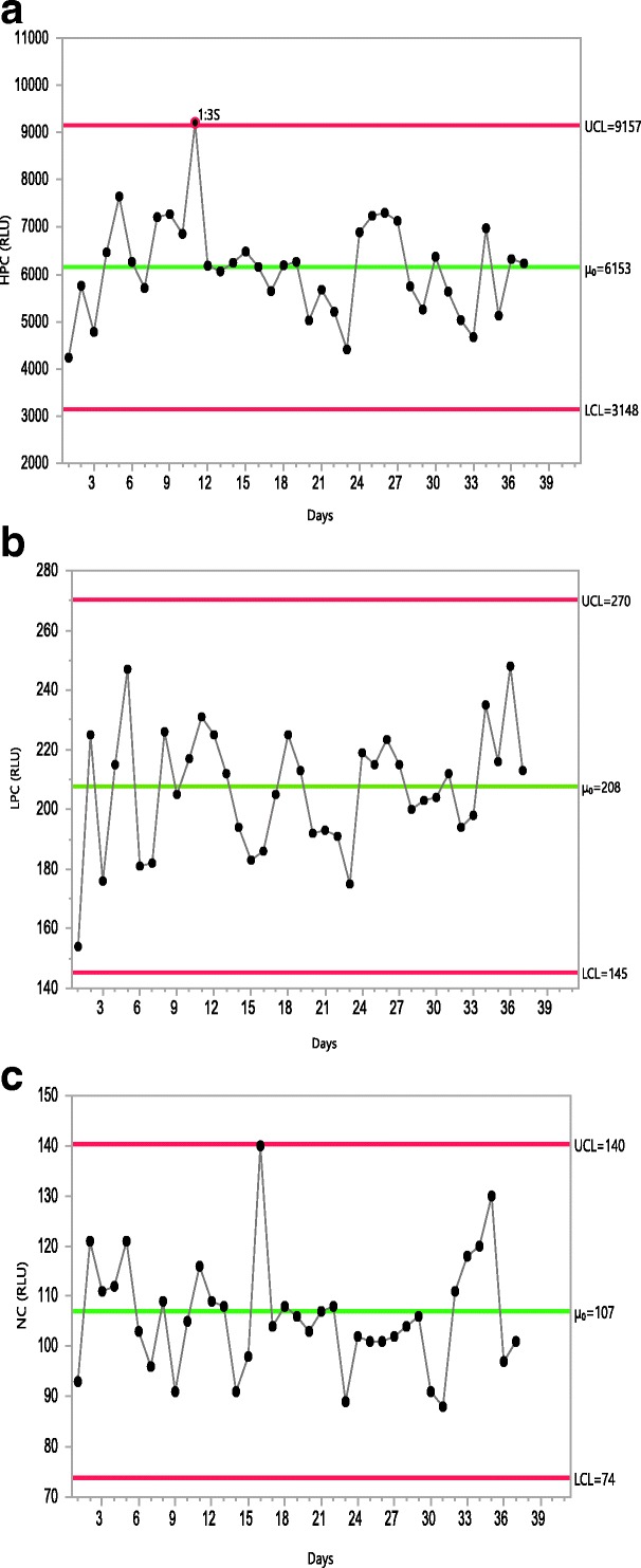
Quality controls (QCs) are the primary indices of assay performance and an important tool in assay lifecycle management. Inclusion of QCs in the testing process allows for the detection of system errors and ongoing assessment of the reliability of the assay. Changes in the performance of QCs are indicative of changes in the assay behavior caused by unintended alterations to reagents or to the operating conditions. The focus of this publication is management of QC life cycle. A consensus view of the ligand binding assay (LBA) community on the best practices for factors that are critical to QC life cycle management including QC preparation, qualification, and trending is presented here.

Keywords: LBA; life cycle management; qualification; quality control; trending.


Figures

Fig. 1
Fig. 1
Example of a Levey-Jennings chart. The plain horizontal red lines are the lower (LCL) and upper (UCL) control limits; the dashed horizontal black line is the observed mean (μ0)
Fig. 2
Fig. 2
Example of a Xbar-R chart. The top panel is the Xbar chart, presenting the mean of each assay; the bottom is the R chart, presenting the observed range within each assay. For both charts, the plain horizontal red lines are the lower and upper control limits; the dashed horizontal black line is the observed mean across assays
Fig. 3
Fig. 3
Westgard detection rules presented all in one plot. The plain black line is the observed mean, pink dashed line is the ± 1 sigma zone, the blue dotted line is the ± 2 sigma zone, and the red dashed line is the ± 3 sigma zone. Each colored marker represents an alert according to the associated rule
Fig. 4
Fig. 4
Predictive distribution of response. The dashed vertical line represents the target and the plain vertical lines represent specification limits. Red zones represent the probability that a future sample result would fall outside specification limits
Fig. 5
Fig. 5
Example of trending of PCs and NC in ADA assays. In this simulated example, the PC and NC performances are trended using Levey-Jennings plots, with UCL and LCL of PCs established as observed mean response (μ0) ± 3 Sigma (while the upper limit of NC was established using mean response + 3 Sigma. In general, NC UCL is critical to restrain the overall background response levels of the assay, while LCL of LPC in some cases could overlap or be below the assay cut point, it is restrained by the assay cut point. Once pre-study method validation has been completed, the UCL and LCL limits are fixed for monitoring the assay performances during in-study sample analysis. In this plot, the controls which exceed their limits have been marked in red circles. For the NC, there is an upward trend in performance by day 16 and again by day 35; this trend was reversed in subsequent runs. Had such trend continued, it would have indicated a drift and would have warranted an investigation. These analyses were performed using JMP software. a HPC (3 Sigma). b LPC (3 Sigma). c NC (3 Sigma)
Fig. 6
Fig. 6
Example of intra- and inter-assay performance trending of three QC levels in a PK assay. Note that in this example, concentrations at each QC level in each assay run were evaluated using %RE against their nominal values. Since two sets of QCs at each level (n = 2) were included in every plate, plotting positional QCs allowed for their comparison and assessment for drift. At each QC level, the open and closed circles represent %RE for the two separate positional QCs on the plate. Red oval highlights the variability between interspersed positional QCs in Run 5
Fig. 7
Fig. 7
Laboratory examples of QC performance trending in biomarker assays. Laboratory QC monitoring data from two different biomarker assays, assay 1 (a, b) and assay 2 (c) analyzed over 987 and 686 days, respectively. a QC values of assay 1 plotted versus the analytical run number. Each plate has two sets of QCs per run and individual QC results are listed consecutively such that the total number of runs is half of what is represented. b A subset of the same data used in a but expressed as SDI values and listed relative to the Run ID. c Measured QC values of assay 2 plotted versus analytical run ID demonstrating shifts in performance due to reagent lot changes (at Run 72) and an assay performance issue (at Run 125). Note: in these examples, QCs from failed plates were included. The plots were performed in Microsoft Excel

References

    1. DeSilva B, Smith W, Weiner R, Kelley M, Smolec J, Lee B, et al. Recommendations for the bioanalytical method validation of ligand-binding assays to support pharmacokinetic assessments of macromolecules. AAPS J. 2003;20(11):1885–1900. - PubMed
    1. Viswanathan CT, Bansal S, Booth B, DeStafano J, Rose MJ, Sailstad J, et al. Quantitative bioanalytical methods validation and implementation: best practices for chromatographic and ligand binding assays. Pharm.Res. 2007;24(10):1962–1973. doi: 10.1007/s11095-007-9291-7. - DOI - PubMed
    1. US Food and Drug Administration . Guidance for industry: bioanalytical method validation. Silver Spring: Center for Drug Evaluation and Research; 2018.
    1. Azadeh M, Gorovits B, Kamerud J, MacMannis S, Safavi A, Sailstad J, Sondag P. Calibration curves in quantitative ligand binding assays: recommendations and best practices for preparation, design, and editing of calibration curves. AAPS J. 2018;20:22. doi: 10.1208/s12248-017-0159-4. - DOI - PubMed
    1. Nowatzke W, Woolf E. Best practices during bioanalytical method validation for the characterization of assay reagents and the evaluation of analyte stability in assay standards, quality controls, and study samples. AAPS J. 2007;9(2):F117–E122. doi: 10.1208/aapsj0902013. - DOI - PMC - PubMed

Publication types

LinkOut - more resources