. 2019 Dec;12(2-3):67-76.
doi: 10.1007/s12307-019-00228-y. Epub 2019 Jul 11.

Pleiotropic Effects of Epithelial Mesenchymal Crosstalk on Head and Neck Cancer: EMT and beyond

Affiliations

Pleiotropic Effects of Epithelial Mesenchymal Crosstalk on Head and Neck Cancer: EMT and beyond

T B Steinbichler et al. Cancer Microenviron. 2019 Dec.

Abstract

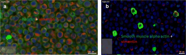
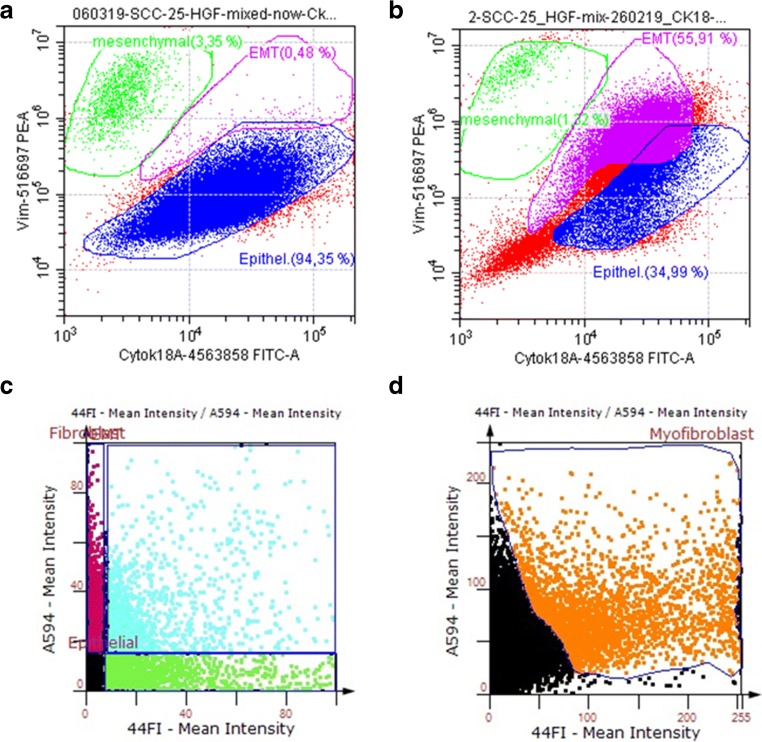
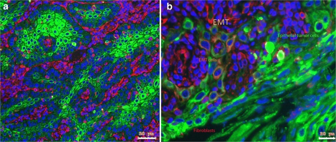
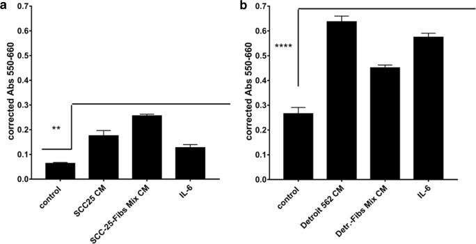
Epithelial mesenchymal crosstalk (EMC) describes the interaction of the tumor stroma and associated fibroblasts with epithelial cancer cells. In this study we analysed the effects of EMC on head and neck cancer cells. In tumor cell lines EMC was induced using media conditioned from a mix-culture of cancer cells and fibroblasts. Cell proliferation and chemotherapy response were assessed using direct cell counting. Flow cytometry, immunohistochemistry of markers of epithelial-mesenchymal transition (EMT) and subsequent TissueFaxs™ acquisition and quantification and western blot analysis were performed. Holotomographic microscopy imaging was used to visualize the effects of EMC on Cisplatin response of SCC-25 cells. EMC induced a hybrid epithelial-mesenchymal phenotype in SCC-25 cells with co-expression of vimentin and cytokeratin. This hybrid phenotype was associated with chemotherapy resistance and increased proliferation of the cells. The EMC conditioned medium led to an activation of the IL-6/STAT3 pathway with subsequent phosphorylation of STAT3. EMC induced a hybrid epithelial-mesenchymal phenotype in HNSCC cells accompanied by increased therapy resistance and cell proliferation. The IL-6/STAT3 pathway might be one of the major pathways involved in these EMC-related effects.

Keywords: EMT; IL-6; JAK/STAT3/Snail signalling; conditioned medium; radiochemotherapy; therapy resistance.


Conflict of interest statement

The authors declare that they have no conflict of interest.

Figures

Fig. 1
Fig. 1
EMC model of HNSCC in cell culture. The mixed culture of SCC-25 cells and HGF fibroblasts functioned as model for EMC. After cell culture and production of conditioned medium (EMC-CM) the cells were embedded in agarose and in paraffin, sectioned and immunostained using anti-pan-cytokeratin (green) and vimentin (red) antibodies (a) or smooth muscle alpha actin (SMA, green) and vimentin (red) antibodies (b). The most abundant component of EMC in cell culture is the EMT cell, showing positive reaction for both pan-cytokeratin and vimentin (coloured in yellow or orange), but SMA+ myofibroblasts (B, green) are also detected in this complex. Bars: 20 μm (n = 5)
Fig. 2
Fig. 2
Flow cytometry and TissueFaxs™/ TissueQuest™ analysis of the EMC model of HNSCC in cell culture. A) SCC-25 oral and HGF fibroblasts were cultured separately and were mixed before flow cytometry. Cells were fixed and stained using the PerFix-nc kit of Beckman Coulter and cytokeratin-18-Alexa Fluor 488, and vimentin- Phycoerythrin direct conjugated antibodies. This sample was used to set the epithelial (blue) and fibroblast (green) gates in the CytoFLEX™ flow cytometer (A). B) If SCC-25 cells and HGF fibroblasts were cultured for production of EMC-CM, the most abundant component of this mixed EMC-culture was the cytokeratin-18–vimentin double positive cell type, which represents the EMT cell (labelled as magenta in panel B). C-D) Agarose and paraffin embedding and subsequent TissueFaxs™ aquisition and TissueQuest™ evaluation of the mix cultured SCC-25 and HGF cells of EMC showed comparable results to B, providing an abundant pan-cytokeratin (detected with Alexa Fluor 488; x-axis on C photographed in 44Fl channel)–vimentin (detected with Alexa Fluor 594; y-axis on C photographed in A594 channel) double positive cell population (light blue on panel C). If the embedded cells were stained with vimentin (detected with Alexa Fluor 594; y-axis on D photographed in A594 channel) and SMA (detected with Alexa Fluor 488; x-axis on D photographed in 44Fl channel); besides the double–negative epithelial cell population and only vimentin–positive fibroblasts (black gate on panel D) a significant vimentin–SMA–double positive cell population (orange gate on panel D) was detectable, which is identified as cancer-associated fibroblast (n = 5)
Fig. 3
Fig. 3
EMC in HNSCC tissue of oral cavity. a) Immunohistochemical reaction of pan-cytokeratin (green) and vimentin (red) in HNSCC tissue. The tumour cell nests (green) are surrounded by fibroblasts (red) and seemed to build functional units in head and neck cancer. b) In several HNSCC samples, this structure was replaced by a mixture of epithelial and mesenchymal cells (B) as pan – cytokeratin – positive epithelial tumour cells (green) and vimentin-positive fibroblasts (red), but also yellow–orange double positive cells, which are mesenchymal transdifferentiated epithelial cells (EMT). Bar represents 50 μm in panel A and 20 μm in panel B (n = 5)
Fig. 4
Fig. 4
EMC increased cell growth in HNSCC cells. SCC-25 (a) and Detroit 562 (b) cells were treated with conditioned medium of single tumor cells, single fibroblasts and mix culture of tumor cells and fibroblasts (EMC). All conditioned media provided a significantly increased cell proliferation compared to the controls measured by cell couting, but in SCC-25 cells the highest effect was observed after treatment with EMC-conditioned medium (EMC-CM; SCC-25-Fibs Mix CM). Using cytokine/chemokine array the most abundant cytokine especially in EMC-CM was interleukin-6 (IL-6), which also supported increased cell growth in both SCC-25 and Detroit 562 cells if used for treatment of the cells at 50 ng/ml. In both panels A and B, the used statistical test was One Way Analysis of Variance (ANOVA) with multiple comparisons of all treatments to controls using Graphpad Prism 7.00 (n = 24)
Fig. 5
Fig. 5
EMC rescues cultured HNSCC cells from chemotherapy induced cell death. As published by us before (4) EMC-conditioned media (EMC-CM) doubled the IC50 value of cisplatin in SCC-25 cells. If SCC-25 cells were treated with cisplatin at IC50 in albumin containing medium for 72 h the cells died and released their adhesion from culture dish (A, B), whereas, the SCC-25 treated with cisplatin at IC50 in EMC CM were alive, metabolically active and large. Furthermore the initiation of multinuclear morphology was observed with several mitochondria and lipid droplets (C, D). A, B, C, D: holotomographic microscopy images of SCC-25 cells using the NanoLIVE system. Scale bars 20 μm
Fig. 6
Fig. 6
EMC-CM transmits induction of EMT through IL-6 - STAT3 signalling. SCC-25 cells were treated with conditioned media of single culture of SCC-25 cells (A, 1), single culture of HGF fibroblasts (A, 2), mixed EMC culture (A, 3), or were indirect co-cultured with HGF fibroblasts (A, 4). A) Western blot analysis of treated SCC-25 for STAT3, phospho-STAT3, Snail and Slug. GAPDH was used as loading control. The SCC-25 cells constitutively synthesized STAT3 and Snail. STAT-3 was only phosphorylated by mixed EMC-CM treatment, which also achieved a significant induction of Slug protein synthesis. B) Western blot analysis of treated SCC-25 cells for STAT3, phospho-STAT3. β-actin was used as loading control. 1: cells cultured in albumin-containing medium, 2: cells treated with CM of single culture of SCC-25 cells, 3: cells treated with CM of single culture of HGF fibroblasts, 4: cells treated with CM of mixed EMC culture, 5: cells treated with 50 ng/ml IL-6, 6: commercial available positive control of induced STAT3 phosphorylation. STAT3 phosphorylation was achieved by CM of mixed EMC culture or by IL-6. C-D) Normalized optical density quantification of western blots presented in B, C: STAT3, D: phospho-STAT3 (n = 3)

References

    1. Hui L, Chen Y. Tumor microenvironment: sanctuary of the devil. Cancer Lett. 2015;368(1):7–13. - PubMed
    1. Metzler VM, Pritz C, Riml A, Romani A, Tuertscher R, Steinbichler T, Dejaco D, Riechelmann H, Dudás J. Separation of cell survival, growth, migration, and mesenchymal transdifferentiation effects of fibroblast secretome on tumor cells of head and neck squamous cell carcinoma. Tumour biology : the journal of the International Society for Oncodevelopmental Biology and Medicine. 2017;39(11):1010428317705507. - PMC - PubMed
    1. Chan R, Sethi P, Jyoti A, McGarry R, Upreti M. Investigating the Radioresistant properties of lung Cancer stem cells in the context of the tumor microenvironment. Radiat Res. 2016;185(2):169–181. - PMC - PubMed
    1. Steinbichler TB, Metzler V, Pritz C, Riechelmann H, Dudas J. Tumor-associated fibroblast-conditioned medium induces CDDP resistance in HNSCC cells. Oncotarget. 2016;7(3):2508–2518. - PMC - PubMed
    1. Steinbichler TB, Alshaimaa A, Maria MV, Daniel D, Herbert R, Jozsef D, et al. Epithelial-mesenchymal crosstalk induces radioresistance in HNSCC cells. Oncotarget. 2018;9(3):3641–3652. - PMC - PubMed