. 2019 Aug;60(8):1627-1638.
doi: 10.1111/epi.16283. Epub 2019 Jul 12.

Identifying the neural basis of a language-impaired phenotype of temporal lobe epilepsy

Affiliations

Identifying the neural basis of a language-impaired phenotype of temporal lobe epilepsy

Erik Kaestner et al. Epilepsia. 2019 Aug.

Abstract

Objective: To identify neuroimaging and clinical biomarkers associated with a language-impaired phenotype in refractory temporal lobe epilepsy (TLE).

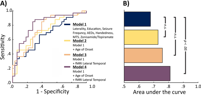
Methods: Eighty-five patients with TLE were characterized as language-impaired (TLE-LI) or non-language-impaired (TLE-NLI) based on comprehensive neuropsychological testing. Structural magnetic resonance imaging (MRI), diffusion tensor imaging, and functional MRI (fMRI) were obtained in patients and 47 healthy controls (HC). fMRI activations and cortical thickness were calculated within language regions of interest, and fractional anisotropy (FA) was calculated within deep white matter tracts associated with language. Analyses of variance were performed to test for differences among the groups in imaging measures. Receiver operator characteristic curves were used to determine how well different clinical versus imaging measures discriminated TLE-LI from TLE-NLI.

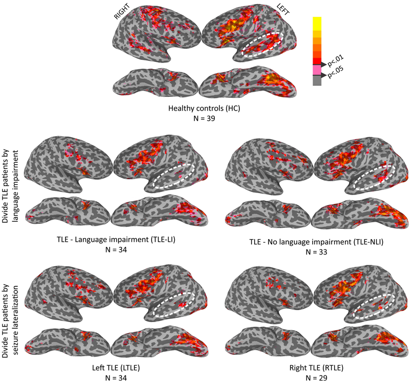
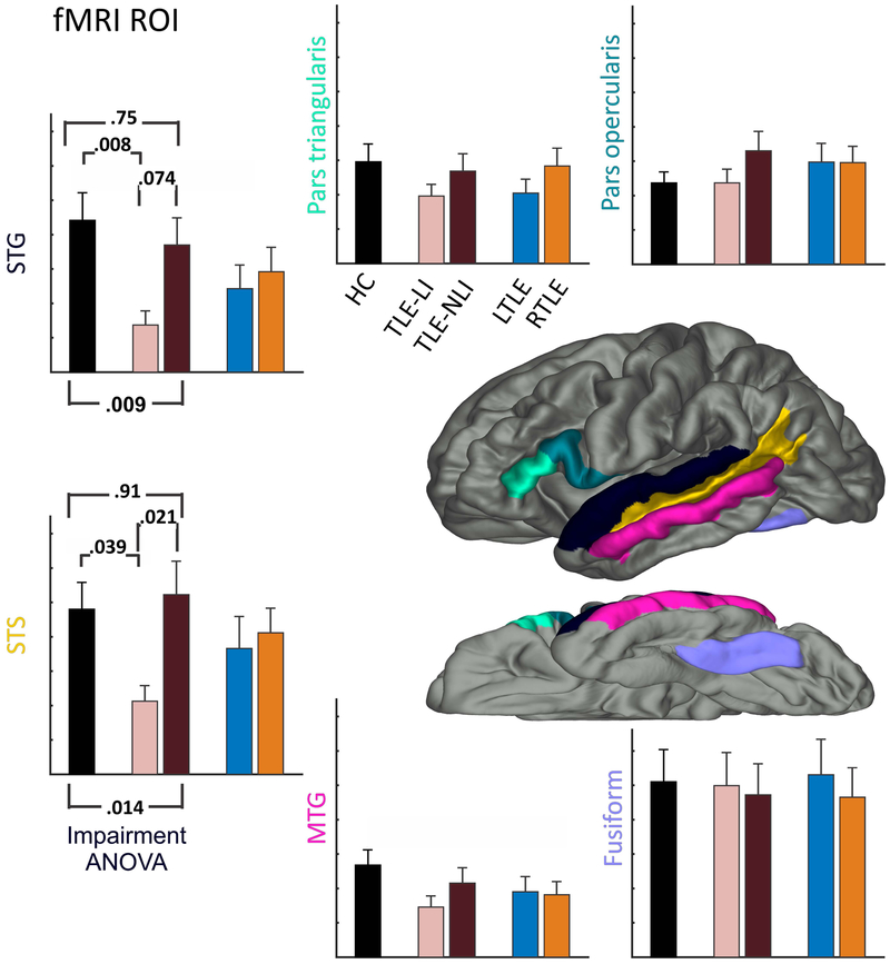
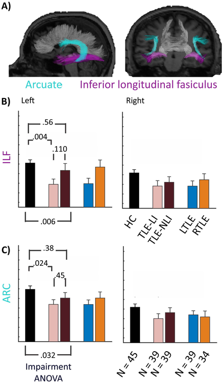
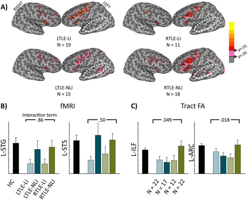
Results: TLE-LI patients showed significantly less activation within left superior temporal cortex compared to HC and TLE-NLI, regardless of side of seizure onset. TLE-LI also showed decreased FA in the inferior longitudinal fasciculus and arcuate fasciculus compared to HC. Cortical thickness did not differ between groups in any region. A model that included language-related fMRI activations within the superior temporal gyrus, age at onset, and demographic variables was the most predictive of language impairment (area under the curve = 0.80).

Significance: These findings demonstrate a unique imaging signature associated with a language-impaired phenotype in TLE, characterized by functional and microstructural alterations within the language network. Reduced left superior temporal activation combined with compromise to language association tracts underlies this phenotype, extending our previous work on cognitive phenotypes that could have implications for treatment-planning or cognitive progression in TLE.

Keywords: clinical biomarkers; cognitive phenotype; diffusion tensor imaging; functional magnetic resonance imaging; neural substrate; neuroimaging.


Conflict of interest statement

Disclosure

None of the authors has any conflict of interest to disclose. We confirm that we have read the Journal’s position on issues involved in ethical publication and affirm that this report is consistent with those guidelines.

Figures

Figure 1.
Figure 1.. fMRI cortical surface maps during a lexical-semantic task demonstrate activation differences in superior temporal regions.
Surface fMRI activation for the new words - false-font contrast for HC (top row), TLE patients split by language impairment (middle row), and TLE patients split by side of seizure onset (bottom row).
Figure 2.
Figure 2.. ROIs confirm that decreases in fMRI activation for language impairment restricted to superior temporal regions.
Six ROIs associated with lexical-semantic processing are displayed on the brain. The surrounding bar graphs represent the number of active voxels in each ROI for the HC (black), followed by TLE split by language impairment into TLE-LI (light green) and TLE-NLI (dark green), then TLE split by side of seizure onset into LTLE (blue) and RTLE (red). Error bars are standard error of the mean. Significant p-values for the Impairment ANOVA are noted, and p-values are reported for follow-up pairwise tests between HC vs TLE-LI and TLE-NLI vs TLE-LI.
Figure 3.
Figure 3.. White matter integrity is compromised in TLE-LI compared to HC in the ILF and ARC.
A) Sagittal and coronal rendering of the arcuate fasciculus (ARC) and inferior longitudinal fasciculus (ILF). B) ILF and C) ARC bar graphs representing the FA of the HC, followed by TLE split by language impairment into TLE-LI (light green) and TLE-NLI (dark green), then TLE split by side of seizure onset into LTLE (blue) and RTLE (red). Error bars are standard error of the mean. Significant p-values for the Impairment ANOVA are noted, and p-values are reported for follow-up pairwise tests between HC vs TLE-LI and TLE-NLI vs TLE-LI.
Figure 4.
Figure 4.. Splitting by side of seizure onset reveals preserved fMRI language-impairment effects but an interaction for tract FA.
A) Surface fMRI activation for the new words - false-font contrast for the 4 TLE groups. B) Number of active voxels in each ROI for HC (black) followed by the TLE split into LTLE-LI (light teal), LTLE-NLI (dark teal), RTLE-LI (light green), and RTLE-NLI (dark green). The p-value is from the interaction term of a 2 (TLE-LI, TLE-NLI) by 2 (LTLE, RTLE) ANOVA. C) Tract FA in each tract, the same as in B).
Figure 5.
Figure 5.. ROC curves and Area Under the Curve comparing model performance when discriminating TLE-LI from TLE-NLI.
A) The ROC curves associated with 4 logistic regression models. Models 2-4 include Model 1 plus additional variable(s). B) The area under the curve associated with each ROC curve. Significance values show whether models 2-4 significantly outperformed model 1 as determined by confidence intervals from 10,000 bootstrapped samples.

References

    1. Bell B, Lin JJ, Seidenberg M, Hermann B. The neurobiology of cognitive disorders in temporal lobe epilepsy. Nat Rev Neurol. 2011;7(3):154–164. doi:10.1038/nrneurol.2011.3 - DOI - PMC - PubMed
    1. Hermann B, Seidenberg M, Lee E-J, Chan F, Rutecki P. Cognitive phenotypes in temporal lobe epilepsy. J Int Neuropsychol Soc 2007;13(01). doi:10.1017/S135561770707004X - DOI - PubMed
    1. Dabbs K, Jones J, Seidenberg M, Hermann B. Neuroanatomical correlates of cognitive phenotypes in temporal lobe epilepsy. Epilepsy Behav. 2009;15(4):445–451. doi:10.1016/j.yebeh.2009.05.012 - DOI - PMC - PubMed
    1. Rodríguez-Cruces R, Velázquez-Pérez L, Rodríguez-Leyva I, et al. Association of white matter diffusion characteristics and cognitive deficits in temporal lobe epilepsy. Epilepsy Behav 2018;79:138–145. doi:10.1016/j.yebeh.2017.11.040 - DOI - PubMed
    1. Reyes A, Kaestner E, Bahrami N, et al. Cognitive phenotypes in temporal lobe epilepsy are associated with distinct patterns of white matter network abnormalities. Neurology. (In Press). - PMC - PubMed

Publication types

MeSH terms