. 2019 Jul 12;15(7):e1007747.
doi: 10.1371/journal.ppat.1007747. eCollection 2019 Jul.

A set of Arabidopsis genes involved in the accommodation of the downy mildew pathogen Hyaloperonospora arabidopsidis

Affiliations

A set of Arabidopsis genes involved in the accommodation of the downy mildew pathogen Hyaloperonospora arabidopsidis

Martina Katharina Ried et al. PLoS Pathog. .

Abstract

The intracellular accommodation structures formed by plant cells to host arbuscular mycorrhiza fungi and biotrophic hyphal pathogens are cytologically similar. Therefore we investigated whether these interactions build on an overlapping genetic framework. In legumes, the malectin-like domain leucine-rich repeat receptor kinase SYMRK, the cation channel POLLUX and members of the nuclear pore NUP107-160 subcomplex are essential for symbiotic signal transduction and arbuscular mycorrhiza development. We identified members of these three groups in Arabidopsis thaliana and explored their impact on the interaction with the oomycete downy mildew pathogen Hyaloperonospora arabidopsidis (Hpa). We report that mutations in the corresponding genes reduced the reproductive success of Hpa as determined by sporangiophore and spore counts. We discovered that a developmental transition of haustorial shape occurred significantly earlier and at higher frequency in the mutants. Analysis of the multiplication of extracellular bacterial pathogens, Hpa-induced cell death or callose accumulation, as well as Hpa- or flg22-induced defence marker gene expression, did not reveal any traces of constitutive or exacerbated defence responses. These findings point towards an overlap between the plant genetic toolboxes involved in the interaction with biotrophic intracellular hyphal symbionts and pathogens in terms of the gene families involved.


Conflict of interest statement

The authors have declared that no competing interests exist.

Figures
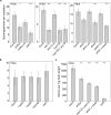
Fig 1
Fig 1. Mutations in A. thaliana SNUPO genes reduce the reproductive success of the oomycete downy mildew pathogen Hpa.
(a + b) Bar charts represent the mean number of sporangiophores ± s.e.m on infected cotyledons of A. thaliana wild-type (Col-0) or the indicated mutants 4 dpi with Hpa isolate Noco2. n = 14–63 (c) Bar charts represent the mean number of spores ± s.e.m per mg fresh weight isolated from A. thaliana wild-type (Col-0) or the indicated mutants 4 dpi with Hpa isolate Noco2. n = 9. Stars indicate significant differences to Col-0 (Wilcoxon-Mann-Whitney test with Bonferroni-Holm correction; ′, p = 0.066 *, p < 0.05; **, p < 0.01; ***, p < 0.001). Each experiment was performed at least three times independently.
Fig 2
Fig 2
The morphology of Hpa haustoria is altered in A. thaliana SNUPO mutants. Boxplots represent the percentage of multilobed haustoria among total haustoria on A. thaliana wild-type (Col-0), the indicated mutants and transgenic homozygous T3 complementation lines (pollux co, pPOLLUX:POLLUX; sec13 co, pSEC13:SEC13; shrk1 co, pUbi:ShRK1; shrk2 co, pUbi:ShRK2; LjSYMRK, pUbi:Lotus japonicus SYMRK-mOrange) 5 dpi (a + b), or 4, 5 and 7 dpi with Hpa isolate Noco2 (d). For each genotype, at least ten independent stretches of hyphae per leaf have been analysed on at least 5 leaves. On average, 1100 haustoria have been analysed per genotype. (c) Representative pictures of intracellular haustoria are displayed. Bars = 25 μm. (e) Upper panel: Light microscopy of Technovit section of A. thaliana wild-type (Col-0) or the shrk1 x shrk2 double mutant 7 dpi with Hpa isolate Noco2. Bars, 10 μm. Middle panel: Confocal light scanning microscopy of leaves of A. thaliana wild-type (Col-0) or the shrk1 x shrk2 double mutant stained with aniline-blue 5 dpi with Hpa isolate Noco2. For every haustorium, the entry point can be identified by the formation of the usually bright callose neck. Bars, 25 μm. Lower panel: Confocal light scanning microscopy of leaves of A. thaliana wild-type (Col-0) expressing RPW8.2-YFP, which localises to the perihaustorial membrane 7 dpi with Hpa isolate Noco2. *, round haustoria; +, multilobed haustoria; arrows, hyphae. Bars, 10 μm. Black circles, data points outside 1.5 IQR of the upper/lower quartile; bold black line, median; box, Interquartile Range (IQR); whiskers, lowest/highest data point within 1.5 IQR of the lower/upper quartile. Stars indicate significant differences to Col-0 (Wilcoxon-Mann-Whitney test with Bonferroni-Holm correction; *, p < 0.05; **, p < 0.01; ***, p < 0.001). Each experiment was performed at least three times independently.
Fig 3
Fig 3. Mutations in A. thaliana SNUPO genes do not impair the reproductive success of the fungal powdery mildew pathogen E. cruciferarum.
Bar charts represent the mean number of conidiophores ± s.e.m on leaves of the indicated mutants relative to the wild-type (Col-0) 5 dpi with E. cruciferarum. At least ten colonies per leaf on at least 6 leaves have been counted. No significant differences to Col-0 were detected (Wilcoxon-Mann-Whitney test with Bonferroni-Holm correction; *, p < 0.05; **, p < 0.01; ***, p < 0.001).
Fig 4
Fig 4. Activation of defence responses in response to Hpa infection is unaltered in A. thaliana SNUPO mutants.
(a) Expression of PR1, ERF1 or PDF1.2a relative to the housekeeping genes TIP41-like and PP2A in mock-treated samples (black circles) or in Hpa-infected samples (empty circles) was determined in three biological replicates for each genotype by qRT-PCR. Stars indicate significant differences to Col-0 (Dunnett’s Test with Bonferroni correction; *, p < 0.05). (b) Boxplots represent the mean intensity of callose deposition in leaves of A. thaliana wild-type (Col-0) or the indicated mutants 4 dpi with Hpa isolate Noco2. n = 27–72. Circles, data points outside 1.5 IQR of the upper/lower quartile; bold black line, median; box, IQR; whiskers, lowest/highest data point within 1.5 IQR of the lower/upper quartile. No significant differences to Col-0 were detected (Wilcoxon-Mann-Whitney test with Bonferroni-Holm correction; *, p < 0.05; **, p < 0.01; ***, p < 0.001). Representative pictures of haustoria-associated callose deposition at the neck band of Hpa haustoria growing on the indicated mutants are shown on the right. Bars, 25 μm.
Fig 5
Fig 5. Cell death responses are unaltered in A. thaliana SNUPO mutants. Differential interference contrast microscopy of A. thaliana wild-type (Col-0) and CSG mutant leaves stained with trypan-blue lactophenol 4 dpi with Hpa isolate Noco2.
First column, non-infected without cell death; second column, non-infected with random cell death; third column, infected with cell death in, or adjacent to, haustoria-containing cells; fourth column, infected without cell death. Bars, 25 μm. Boxplots represent the number of random (upper graph) or Hpa-associated (lower graph) cell death spots per leaf on A. thaliana wild-type (Col-0) or the indicated mutants 5 dpi with Hpa isolate Noco2. On each leaf four to ten spots have been analysed and a total of five leaves per genotype were investigated. Circles, data points outside 1.5 IQR of the upper/lower quartile; bold black line, median; box, IQR; whiskers, lowest/highest data point within 1.5 IQR of the lower/upper quartile. Stars indicate significant differences to Col-0 (Wilcoxon-Mann-Whitney test with Bonferroni-Holm correction; *, p < 0.05; **, p < 0.01; ***, p < 0.001).
Fig 6
Fig 6. Expression of flg22-inducible genes as well as growth of the bacterial pathogen Pseudomonas syringae are not affected in A. thaliana SNUPO mutants.
(a) Expression of FRK1 and ERF1 relative to the housekeeping genes TIP41-like and PP2A in mock-treated samples (black circles) or in flg22-treated samples (empty circles) was determined in three biological replicates for each genotype by qRT-PCR. The fls2 mutant served as negative control. Stars indicate significant differences to Col-0 (Dunnett’s Test with Bonferroni correction; *, p < 0.01; **, p < 0.001). (b) Bacterial density of Pto DC3000 or Pto DC3000 ΔAvrPto/PtoB on A. thaliana wild-type (Col-0) or the indicated mutants was determined as colony forming units per cm2 2 dpi. Data represent means ± s.d. of six replicate measurements/genotype/data point. Results from one representative of at least four independent experiments are shown.

Similar articles

Cited by

References

    1. Gutjahr C, Parniske M. Cell and developmental biology of arbuscular mycorrhiza symbiosis. Annu Rev Cell Dev Biol. 2013;29: 593–617. 10.1146/annurev-cellbio-101512-122413 - DOI - PubMed
    1. Genre A, Chabaud M, Faccio A, Barker DG, Bonfante P. Prepenetration apparatus assembly precedes and predicts the colonization patterns of arbuscular mycorrhizal fungi within the root cortex of both Medicago truncatula and Daucus carota. Plant Cell. 2008;20: 1407–1420. 10.1105/tpc.108.059014 - DOI - PMC - PubMed
    1. Genre A, Chabaud M, Timmers T, Bonfante P, Barker DG. Arbuscular mycorrhizal fungi elicit a novel intracellular apparatus in Medicago truncatula root epidermal cells before infection. Plant Cell. 2005;17: 3489–3499. 10.1105/tpc.105.035410 - DOI - PMC - PubMed
    1. Parniske M. Intracellular accommodation of microbes by plants: A common developmental program for symbiosis and disease? Current Opinion in Plant Biology. 2000. pp. 320–328. 10.1016/S1369-5266(00)00088-1 - DOI - PubMed
    1. Hückelhoven R, Panstruga R. Cell biology of the plant-powdery mildew interaction. Current Opinion in Plant Biology. 2011. pp. 738–746. 10.1016/j.pbi.2011.08.002 - DOI - PubMed

Publication types

LinkOut - more resources