

Development and Cell Biology of the Blood-Brain Barrier
- PMID: 31299172
- PMCID: PMC8934576
- DOI: 10.1146/annurev-cellbio-100617-062608
Development and Cell Biology of the Blood-Brain Barrier
Abstract
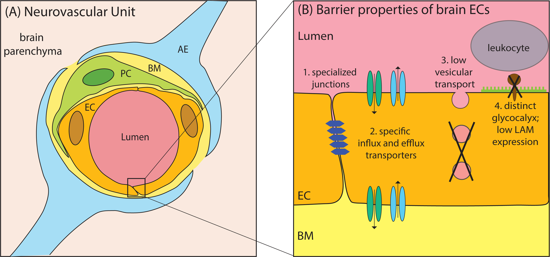
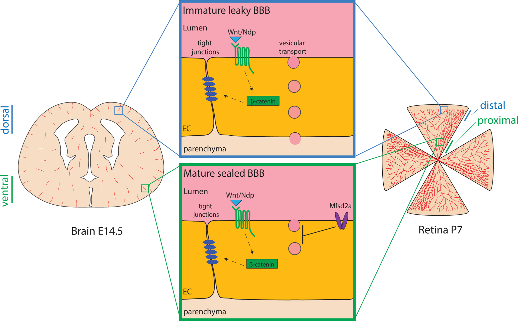
The vertebrate vasculature displays high organotypic specialization, with the structure and function of blood vessels catering to the specific needs of each tissue. A unique feature of the central nervous system (CNS) vasculature is the blood-brain barrier (BBB). The BBB regulates substance influx and efflux to maintain a homeostatic environment for proper brain function. Here, we review the development and cell biology of the BBB, focusing on the cellular and molecular regulation of barrier formation and the maintenance of the BBB through adulthood. We summarize unique features of CNS endothelial cells and highlight recent progress in and general principles of barrier regulation. Finally, we illustrate why a mechanistic understanding of the development and maintenance of the BBB could provide novel therapeutic opportunities for CNS drug delivery.
Keywords: astrocytes; blood-brain barrier; central nervous system; endothelial cells; neurovascular unit; pericytes.
Figures
References
-
- Alvarez JI, Dodelet-Devillers A, Kebir H, Ifergan I, Fabre PJ, et al. 2011. The Hedgehog pathway promotes blood-brain barrier integrity and CNS immune quiescence. Science 334: 1727–31 - PubMed
-
- Armulik A, Genove G, Mae M, Nisancioglu MH, Wallgard E, et al. 2010. Pericytes regulate the blood-brain barrier. Nature 468: 557–61 - PubMed
-
- Armulik A, Mae M, Betsholtz C. 2011. Pericytes and the blood-brain barrier: recent advances and implications for the delivery of CNS therapy. Ther Deliv 2: 419–22 - PubMed
Publication types
MeSH terms
Grants and funding
LinkOut - more resources
Full Text Sources
Other Literature Sources
