. 2019 Sep:84:106180.
doi: 10.1016/j.leukres.2019.106180. Epub 2019 Jul 3.

Interleukin-8 blockade prevents activated endothelial cell mediated proliferation and chemoresistance of acute myeloid leukemia

Affiliations

Interleukin-8 blockade prevents activated endothelial cell mediated proliferation and chemoresistance of acute myeloid leukemia

Vindhya Vijay et al. Leuk Res. 2019 Sep.

Abstract

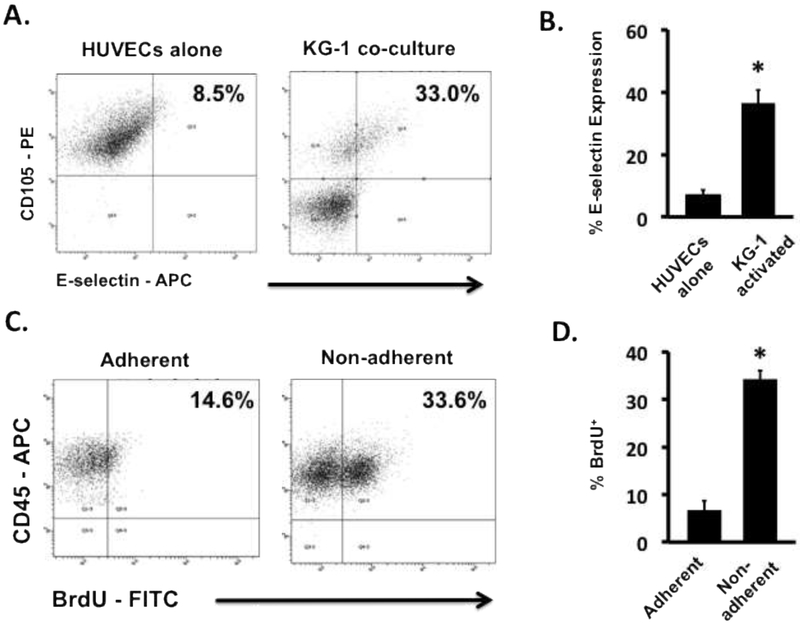
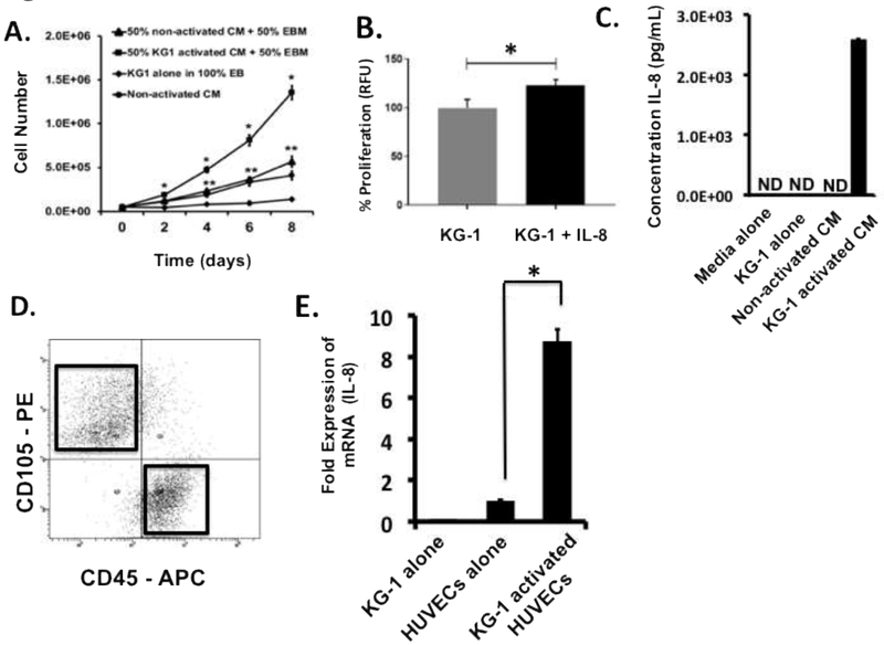
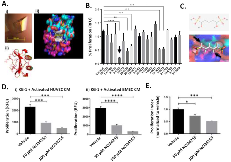
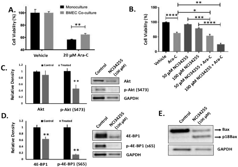
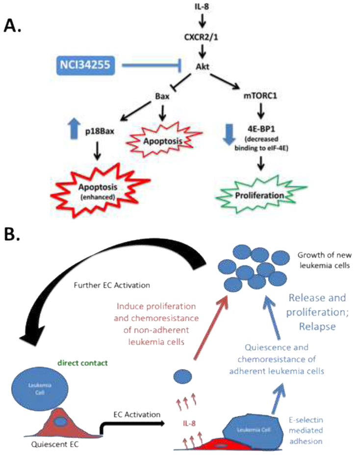
One of the greatest challenges in treating acute myeloid leukemia (AML) is chemotherapy refractory disease. Previously, we demonstrated a novel mechanism whereby AML-induced endothelial cell (EC) activation leads to subsequent leukemia cell adherence, quiescence and chemoresistance, identifying activated ECs as potential mediators of relapse. We now show mechanistically that EC activation induces the secretion of interleukin-8 (IL-8) leading to significant expansion of non-adherent AML cells and resistance to cytarabine (Ara-C). Through crystallography and computational modeling, we identified a pocket within IL-8 responsible for receptor binding, screened for small molecules that fit within this pocket, and blocked IL-8 induced proliferation and chemo-protection of AML cells with a hit compound. Results from this study show a new therapeutic strategy for targeting the sanctuary of an activated leukemia microenvironment.

Keywords: Acute myeloid leukemia; Chemoresistance; Endothelial cell; Interleukin-8; Microenvironment; Vascular niche.


Figures

Figure 1.
Figure 1.. Leukemia cells activate resting ECs resulting in altered leukemia cell proliferation.
(A) Representative flow cytometry plots showing E-selectin levels on KG-1 activated HUVECs and non-activated HUVEC controls. (B) The levels of E-selectin expression on the surface of ECs showed significant increases when activated with KG-1 cells in co-culture. * p < 0.05 (C) Representative flow cytometry plots showing BrdU uptake by adherent and non-adherent AML cells in contact co-cultures of HUVECs and KG-1 cells. (D) BrdU uptake in non-adherent KG-1 cell populations was significantly higher in comparison to adherent populations indicating a proliferative phenotype. * p < 0.05
Figure 2.
Figure 2.. Leukemia activated ECs secrete IL-8 which enhances leukemia cell expansion.
(A) Growth curves of KG-1 cells grown in different media are shown. Supplementing base EBM media with 50% (by volume) activated CM induces significant levels of KG-1 cell growth in comparison to all other cultures tested including those supplemented with non-activated CM. Cells from each culture cohort were enumerated every 2-days over an 8-day culture period. * p < 0.05 versus all other cultures; ** p < 0.05 versus 100% EBM cultures. (B) KG-1 proliferation was significantly enhanced when media was supplemented with IL-8. (C) Fresh, unfrozen supernatants from non-activated and activated co-cultures were evaluated for the production of IL-8 by ELISA. Higher IL-8 concentrations were observed in activated CM. As controls, supernatants from KG-1 alone cultures and pure EGM-2 media were analyzed. Values were extrapolated from standard curves with linear detection limits of 10–3300 pg/mL. ND indicates non-detectable levels. (D) Flow cytometry-based sorting was used to isolate ECs from activating co-cultures. Flow cytometry plots identify gates established for sorting. Representative plots are shown. ECs were isolated based on CD105 (PE) expression while KG-1 cells were identified using CD45 (APC). (E) Sorted ECs were analyzed for mRNA expression levels for IL-8 using qRT-PCR. Non-activated ECs and KG-1 cells alone were analyzed as negative controls. * p < 0.05 versus ECs alone
Figure 3.
Figure 3.. Identification of small molecule inhibitor of IL-8 that can significantly reduce leukemia cell expansion.
(A) Representative IL-8 crystal grown using the vapor diffusion hanging drop method. IL8 crystal yielding complete X-ray diffraction data set to 0.95 Å grown in 0.17 M Ammonium acetate, 0.085 M Sodium citrate tribasic dihydrate pH 5.6, 20% w/v Polyethylene glycol 4,000 and 15% v/v Glycerol (i). The IL-8 dimer is shown as a ribbon diagram in red. Side chains are shown for residues implicated in receptor binding based on mutagenesis studies (ii). The molecular surface of the IL-8 dimer is shown with blue for nitrogen, red for oxygen, cyan for carbon in the (top IL-8 subunit) and magenta for carbon (bottom IL-8 subunit). Residues implicated in receptor binding are shown in white. The scoring grid for molecular docking is shown as a box. 139,735 NCI/DTP compounds were docked into the binding site in silico to predict the binding affinity for IL-8 (iii). (B) Docking studies identified 19 small molecules capable of binding IL-8’s receptor binding site. Treatment of KG-1 cells with these molecules identified 5 that were able to significantly decrease KG-1 proliferation. The best hit molecule is identified with an arrow. ** p < 0.01; *** p < 0.001; **** p < 0.0001 (C) The best hit molecule was NCI34255 which interferes with binding at the homodimeric interface of two IL-8 sub-units (see arrows). (D) KG-1 cells were grown in CM from KG-1 activated ECs (HUVECs and BMECs) supplemented with NCI34255. The presence of NCI34255 was able to significantly decrease IL-8 induced cell proliferation contrary to the enhanced proliferation observed in previous CM studies. *** p < 0.001; **** p < 0.0001 (E) Supplementation of co-cultures comprising KG-1/BMECs with NCI34255 showed significant decreases in non-adherent KG-1 cell proliferation indicating the ability of NCI34255 to overcome activated EC-generated IL-8 signaling. * p < 0.05; *** p < 0.001
Figure 4.
Figure 4.. IL-8 induces chemoresistance which is abrogated using NCI34255.
(A) The response of KG-1 cells in monoculture or non-adherent KG-1 cells in co-culture to Ara-C was tested and showed that KG-1 cells are significantly less sensitive to Ara-C when cultured with ECs. ** p < 0.01 (B) Co-cultures were treated with Ara-C, NCI34255 or a combination of both. Significant decreases in cell viability was observed when Ara-C alone treated groups were compared to Ara-C + NCI34255 treated groups demonstrating that NCI34255 was able to augment Ara-C and enhance apoptotic responses. NCI34255 alone treated groups did affect cell viability in comparison to vehicle controls. * p < 0.05; ** p < 0.01; *** p < 0.001; **** p < 0.0001 (C, D) Analysis of Akt activity in non-adherent KG-1 cells collected from EC co-culture treated with NCI34255 showed significantly decreased levels of phosphorylated Akt in comparison to non-treated controls (C). A similar analysis also showed a significant decrease in phosphorylated 4E-BP1 a downstream target of mTORC1 in the same cell population (D). ** p < 0.01 (E) Bax expression in KG-1 cells isolated from co-cultures exposed to NCI34255 treatment was assessed. The results showed that Bax is cleaved in response to NCI34255 forming the apoptosis enhancing truncated form, p18Bax.
Figure 5.
Figure 5.
(A) Schematic summarizing observed effects of NCI34255 on Akt induced proliferation and survival of AML cells. Treatment with NCI34255 reduces the effects of EC activation through Akt signaling (blue lines) resulting in decreased proliferation and enhanced apoptosis (in the presence of Ara-C) of AML cells. (B) The impact of EC activation on adherent and non-adherent AML cell populations is shown. Adherent cells become quiescent and chemoresistant implicating them in relapse. Non-adherent cells show significant expansion and chemoresistance following EC activation identifying them as the cellular source for enhanced leukemia cellularity. Therapies aimed at targeting this process may provide new avenues for the optimal treatment of patients with AML.

Similar articles

Cited by

References

    1. Tallman MS, Gilliland DG, Rowe JM, Drug therapy for acute myeloid leukemia, Blood 106(4) (2005) 1154–63. - PubMed
    1. Guerrouahen BS, Al-Hijji I, Tabrizi AR, Osteoblastic and vascular endothelial niches, their control on normal hematopoietic stem cells, and their consequences on the development of leukemia, Stem Cells Int 2011 (2011) 375857. - PMC - PubMed
    1. Doan PL, Chute JP, The vascular niche: home for normal and malignant hematopoietic stem cells, Leukemia 26(1) (2012) 54–62. - PubMed
    1. Kiel MJ, Radice GL, Morrison SJ, Lack of evidence that hematopoietic stem cells depend on N-cadherin-mediated adhesion to osteoblasts for their maintenance, Cell Stem Cell 1(2) (2007) 204–17. - PubMed
    1. Cogle CR, Goldman DC, Madlambayan GJ, Leon RP, Al Masri A, Clark HA, Asbaghi SA, Tyner JW, Dunlap J, Fan G, Kovacsovics T, Liu Q, Meacham A, Hamlin KL, Hromas RA, Scott EW, Fleming WH, Functional integration of acute myeloid leukemia into the vascular niche, Leukemia 28(10) (2014) 1978–87. - PMC - PubMed

Publication types

MeSH terms