. 2019 Jul 12;11(1):102.
doi: 10.1186/s13148-019-0696-z.

Apabetalone (RVX-208) reduces vascular inflammation in vitro and in CVD patients by a BET-dependent epigenetic mechanism

Affiliations

Apabetalone (RVX-208) reduces vascular inflammation in vitro and in CVD patients by a BET-dependent epigenetic mechanism

Laura M Tsujikawa et al. Clin Epigenetics. .

Abstract

Background: Apabetalone (RVX-208) is a bromodomain and extraterminal protein inhibitor (BETi) that in phase II trials reduced the relative risk (RR) of major adverse cardiac events (MACE) in patients with cardiovascular disease (CVD) by 44% and in diabetic CVD patients by 57% on top of statins. A phase III trial, BETonMACE, is currently assessing apabetalone's ability to reduce MACE in statin-treated post-acute coronary syndrome type 2 diabetic CVD patients with low high-density lipoprotein C. The leading cause of MACE is atherosclerosis, driven by dysfunctional lipid metabolism and chronic vascular inflammation (VI). In vitro studies have implicated the BET protein BRD4 as an epigenetic driver of inflammation and atherogenesis, suggesting that BETi may be clinically effective in combating VI. Here, we assessed apabetalone's ability to regulate inflammation-driven gene expression and cell adhesion in vitro and investigated the mechanism by which apabetalone suppresses expression. The clinical impact of apabetalone on mediators of VI was assessed with proteomic analysis of phase II CVD patient plasma.

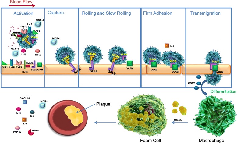
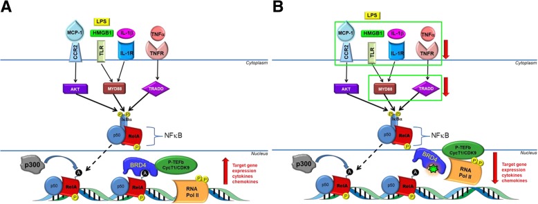
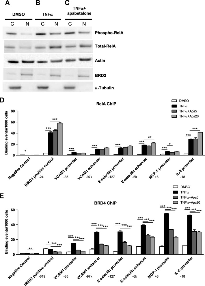
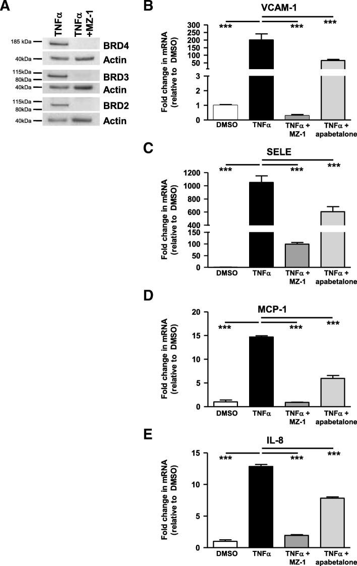
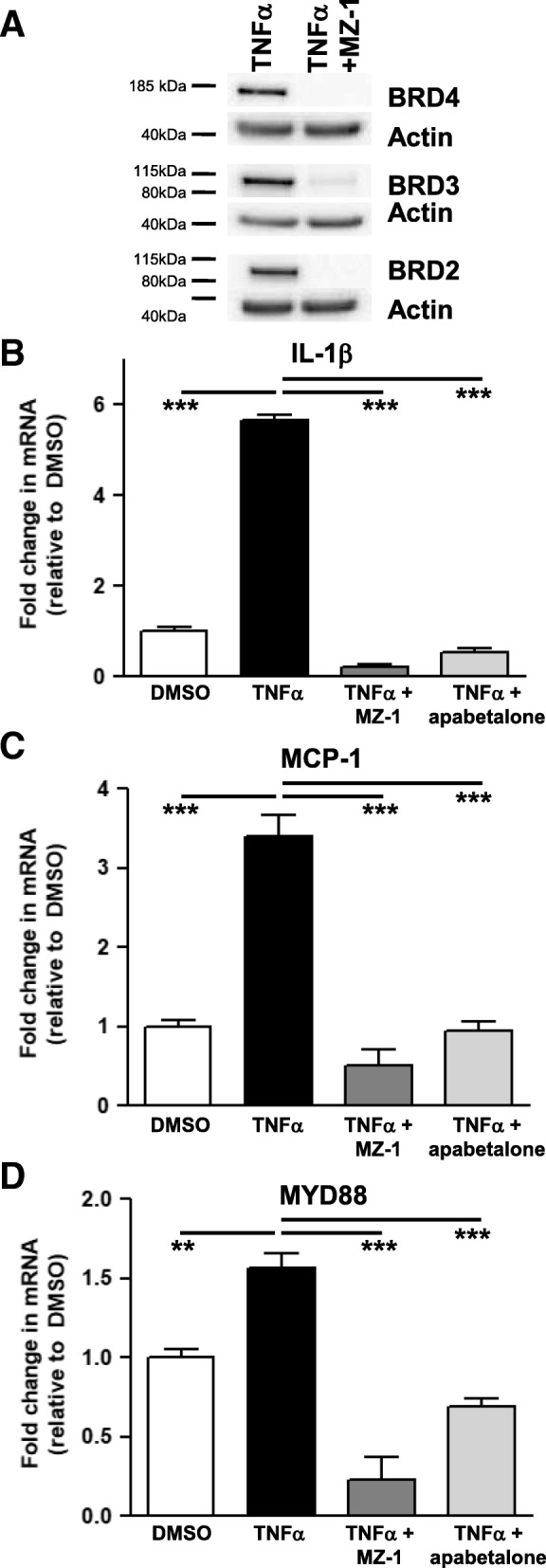
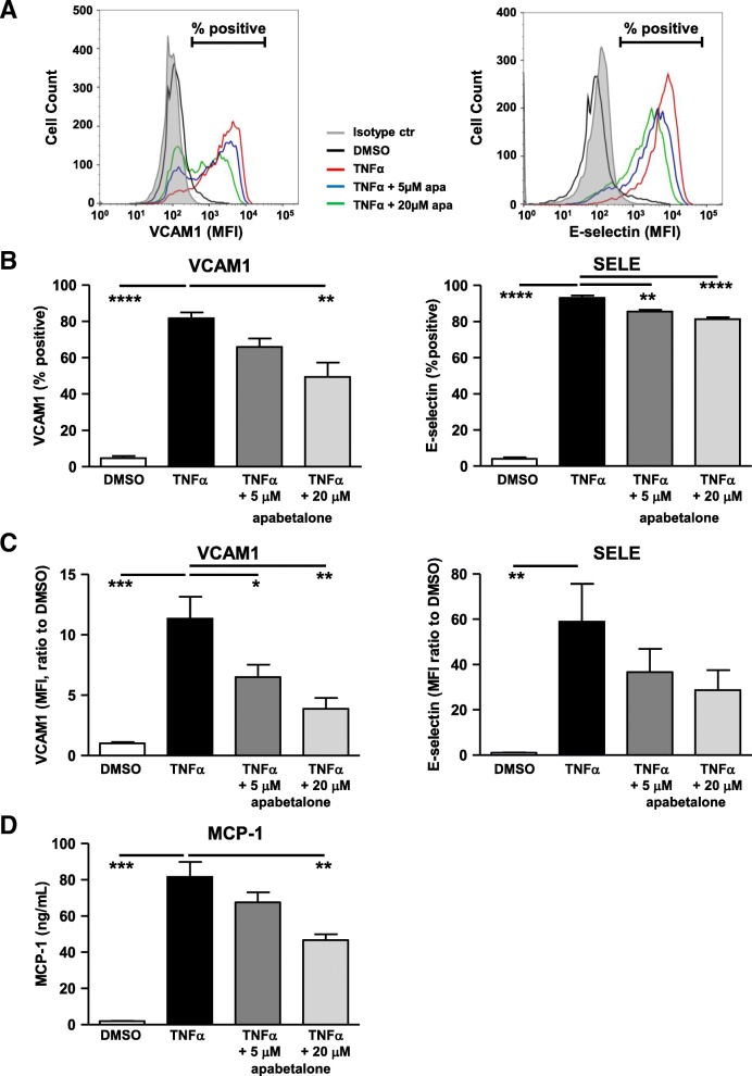
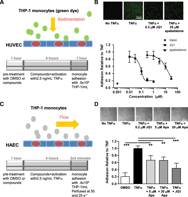
Results: In vitro, apabetalone prevented inflammatory (TNFα, LPS, or IL-1β) induction of key factors that drive endothelial activation, monocyte recruitment, adhesion, and plaque destabilization. BRD4 abundance on inflammatory and adhesion gene promoters and enhancers was reduced by apabetalone. BRD2-4 degradation by MZ-1 also prevented TNFα-induced transcription of monocyte and endothelial cell adhesion molecules and inflammatory mediators, confirming BET-dependent regulation. Transcriptional regulation by apabetalone translated into a reduction in monocyte adhesion to an endothelial monolayer. In a phase II trial, apabetalone treatment reduced the abundance of multiple VI mediators in the plasma of CVD patients (SOMAscan® 1.3 k). These proteins correlate with CVD risk and include adhesion molecules, cytokines, and metalloproteinases. Ingenuity® Pathway Analysis (IPA®) predicted that apabetalone inhibits pro-atherogenic regulators and pathways and prevents disease states arising from leukocyte recruitment.

Conclusions: Apabetalone suppressed gene expression of VI mediators in monocytes and endothelial cells by inhibiting BET-dependent transcription induced by multiple inflammatory stimuli. In CVD patients, apabetalone treatment reduced circulating levels of VI mediators, an outcome conducive with atherosclerotic plaque stabilization and MACE reduction. Inhibition of inflammatory and adhesion molecule gene expression by apabetalone is predicted to contribute to MACE reduction in the phase III BETonMACE trial.

Keywords: Adhesion; Apabetalone; Atherosclerosis; BRD4; Bromodomain; CVD; Diabetes; Epigenetics; HUVEC; THP-1 monocytes; Vascular inflammation; endothelium.


Conflict of interest statement

All authors that are Resverlogix employees receive salaries and shares from the company.

Figures

Fig. 1
Fig. 1
The multi-step process of atherogenesis: Activation of the endothelium, monocyte activation and recruitment, leukocyte capture, rolling, adhesion, firm adhesion, macrophage differentiation, plaque development and stability. Apabetalone downregulates the transcription of each protein labeled in the illustration, thus impacting each step of atherogenesis. At the plaque, circulating and local cytokine expression from endothelial cells and monocytes are downregulated by apabetalone. Activation panel: pink speckles represent multiple cytokine secretion
Fig. 2
Fig. 2
Convergent inflammatory signaling through NF-κB potentiates BRD4-dependent transcription of VI mediators, a result suppressed by apabetalone. a MCP-1, LPS, IL-1β, and TNFα all signal through NF-κB. The stimulants MCP-1, LPS, IL-1β, and TNFα activate their cognate receptors CCR2, TLR, IL-1R, and TNFR respectively. The receptors translate the signal through AKT, MYD88 and TRADD, phosphorylating NF-κB (yellow “p” circles) and releasing RelA-p50 subunits from IκBα. RelA translocates to the nucleus where it binds to consensus DNA binding sequences and is acetylated at K310 by p300 (black “a” circles). BRD4 recognizes and binds to these acetylation marks, recruiting pTEFb to activate RNA Pol II to drive inflammatory gene expression (cytokines, chemokines, and adhesion molecules). b Apabetalone (green 7-point star) competitively inhibits BRD4 BD2 interactions with acetylated lysine marks on RelA. This prevents pTEFb recruitment and Pol II activation, inhibiting the transcription of VI mediators and components of the NF-κB pathway. Green boxes and red arrows indicate genes in the illustration whose expression is reduced by apabetalone
Fig. 3
Fig. 3
Apabetalone does not interfere with NF-kB translocation from the cytoplasm to the nucleus or association of RelA with the chromatin shown via western blot and ChIP. a Western blot: Phospho-RelA and total-RelA is found almost exclusively in the HUVEC cytoplasm (C) under unstimulated conditions (DMSO). b TNFα stimulation induces phospho-RelA and total-RelA translocation to the nucleus (N). c Apabetalone (20 μM) co-treatment (2 h) does not alter translocation. ac The loading control used was β-actin, the nuclear protein control was BRD2 and cytoplasmic control was α-tubulin. d ChIP: RelA occupancy on the VCAM1 enhancer and promoter, the SELE enhancer and promoter, and the promoters of MCP-1 and IL-8 increases substantially with TNFα stimulation. Apabetalone (5 and 20 μM) does not reduce RelA occupancy. e BRD4 occupancy on the VCAM1 enhancer and promoter, the SELE enhancer and promoter, and the promoters of MCP-1 and IL-8 also increases substantially with TNFα stimulation. Apabetalone (5 and 20 μM) diminishes BRD4 occupancy at each of these sites. ChIP locations from transcriptional start sites are indicated by the target gene. Statistical significance was determined through 1-way ANOVA analysis followed by Dunnett’s Multiple Comparison Test using TNFα response for the comparison, where *p < 0.05, **p < 0.01, ***p < 0.001
Fig. 4
Fig. 4
In endothelial cells, MZ-1 and apabetalone prevent TNFα induction of key inflammatory and adhesion marker transcripts through the degradation or inhibition of BET proteins respectively. a MZ-1 (1 μM; 6 h) degrades HUVEC BRD2, BRD3, and BRD4 as shown by western blot. be TNFα stimulation (2 h) fails to induce HUVEC transcription of VCAM-1 (b), MCP-1 (d), and IL-8 (e) following MZ-1 pretreatment (4 + 2 h). SELE (c) induction is reduced but not eliminated. Apabetalone (20 μM) pretreatment (4 + 2 h) also decreases the level of inductions. Statistical significance was determined through 1-way ANOVA analysis followed by Dunnett’s Multiple Comparison Test using TNFα response for the comparison, where *p < 0.05, **p < 0.01, ***p < 0.001
Fig. 5
Fig. 5
In THP-1 cells, MZ-1 and apabetalone prevent TNFα induction of key inflammatory and adhesion marker transcripts. a MZ-1 (6 h; 1 μM) degrades THP-1 BRD2, BRD3, and BRD4 as shown by western blot. bd TNFα stimulation (2 h) fails to induce transcription of IL-1β (b), MCP-1 (c), or MYD88 (d) following MZ-1 pretreatment (6 h). Apabetalone (20 μM) pretreatment (6 h) decreases the transcripts of these genes. Statistical significance was determined through 1-way ANOVA analysis followed by Dunnett’s Multiple Comparison Test using TNFα response for the comparison, where *p < 0.05, **p < 0.01, ***p < 0.001
Fig. 6
Fig. 6
In HUVECs, apabetalone regulation of transcription reduces the abundance of VCAM-1 and MCP-1 proteins. HUVEC cells were stimulated with TNFα and co-treated with apabetalone for 4 h. The surface abundance of VCAM-1 (FITC-CD106) and SELE (APC-CD62E) were measured by flow cytometry. a Representative histogram overlay of HUVEC surface staining for VCAM-1 and SELE. Smaller peaks (% positive reduction) and left-ward curve shifts (MFI reduction) are both indications that there is a reduction in surface expression for the given protein. b Average of % positive cells expressing VCAM1 or SELE on the cell surface relative to the isotype control (the filled gray histogram as indicated in A). c Average mean fluorescent intensity (MFI) of VCAM1 and SELE on HUVEC surface relative to DMSO control. d HUVEC MCP-1 secretion is induced by overnight TNFα stimulation. Co-application with 20 μM apabetalone significantly reduces MCP-1 secretion (BDTM cytometric bead array). In bd, the results represent the mean of four independent experiments ± standard error. Statistical significance was determined through 1-way ANOVA analysis followed by Dunnett’s Multiple Comparison Test using TNFα response for the comparison, where *p < 0.05, **p < 0.01, ***p < 0.001
Fig. 7
Fig. 7
Apabetalone prevents monocyte adhesion to endothelial cells. a Static assay. Endothelial cell monolayer was pretreated with DMSO, JQ1, or apabetalone for 1 h before the addition of TNFα (2.5 ng/ml; 4 h incubation). Monocytes (0.5 × 10⁶cells/mL; loaded with calcein-AM) had 1 h to adhere to the monolayer before washes and fluorescence measures (plate reader). b Upper panel: fluorescent micrographs of monocyte adhesion to endothelial cells under static conditions. Lower panel: dose-response curves titrating JQ1 and apabetalone effect on monocyte /endothelial cell adhesion. JQ1 IC50 = 0.08 μM. apabetalone IC50 = 22 μM. c Flow assay. Pretreatment as in a; monocytes (0.4 × 10⁶ cells/ml) were perfused over the treated monolayer for 3 min with a flow rate of 50 s−1 then for another 3 min with a flow rate of 25 s−1. A high flow rate of 120 s−1 was applied to remove all unbound THP-1 cells, and images were acquired for analysis. d Upper panel: phase-contrast micrographs of monocyte adhesion to endothelial cells under flow conditions. Lower panel: apabetalone pretreatment prevents monocyte adhesion to endothelial cells under flow conditions; 0.2 μM JQ1 and 5 μM apabetalone had a comparable effect on adhesion. Statistical significance was determined through 1-way ANOVA analysis followed by Dunnett’s Multiple Comparison Test using TNFα response for the comparison, where *p < 0.05, **p < 0.01, ***p < 0.001

References

    1. Mestas J, Ley K. Monocyte-endothelial cell interactions in the development of atherosclerosis. Trends Cardiovasc Med. 2008;18:228–232. doi: 10.1016/j.tcm.2008.11.004. - DOI - PMC - PubMed
    1. Tajbakhsh A, Rezaee M, Kovanen PT, Sahebkar A. Efferocytosis in atherosclerotic lesions: Malfunctioning regulatory pathways and control mechanisms. Pharmacol Ther. 2018;188:12–25. doi: 10.1016/j.pharmthera.2018.02.003. - DOI - PubMed
    1. Chistiakov DA, Melnichenko AA, Myasoedova VA, Grechko AV, Orekhov AN. Mechanisms of foam cell formation in atherosclerosis. J Mol Med (Berl). 2017;95:1153–1165. doi: 10.1007/s00109-017-1575-8. - DOI - PubMed
    1. Stefanadis C, Antoniou CK, Tsiachris D, Pietri P. Coronary atherosclerotic vulnerable plaque: current perspectives. J Am Heart Assoc. 2017;6. 10.1161/JAHA.117.005543. - PMC - PubMed
    1. World Heath Organization. Cardiovascular diseases (CVDs) [Webpage]. 2017. https://www.who.int/en/news-room/fact-sheets/detail/cardiovascular-disea.... [updated May 17, 2017; cited 2019 January 10, 2019].

Publication types

MeSH terms