. 2019 Jul 3;12(7):dmm039073.
doi: 10.1242/dmm.039073.

RDH10 function is necessary for spontaneous fetal mouth movement that facilitates palate shelf elevation

Affiliations

RDH10 function is necessary for spontaneous fetal mouth movement that facilitates palate shelf elevation

Regina M Friedl et al. Dis Model Mech. .

Abstract

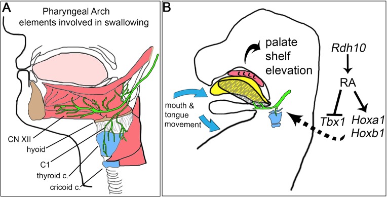
Cleft palate is a common birth defect, occurring in approximately 1 in 1000 live births worldwide. Known etiological mechanisms of cleft palate include defects within developing palate shelf tissues, defects in mandibular growth and defects in spontaneous fetal mouth movement. Until now, experimental studies directly documenting fetal mouth immobility as an underlying cause of cleft palate have been limited to models lacking neurotransmission. This study extends the range of anomalies directly demonstrated to have fetal mouth movement defects correlated with cleft palate. Here, we show that mouse embryos deficient in retinoic acid (RA) have mispatterned pharyngeal nerves and skeletal elements that block spontaneous fetal mouth movement in utero Using X-ray microtomography, in utero ultrasound video, ex vivo culture and tissue staining, we demonstrate that proper retinoid signaling and pharyngeal patterning are crucial for the fetal mouth movement needed for palate formation. Embryos with deficient retinoid signaling were generated by stage-specific inactivation of retinol dehydrogenase 10 (Rdh10), a gene crucial for the production of RA during embryogenesis. The finding that cleft palate in retinoid deficiency results from a lack of fetal mouth movement might help elucidate cleft palate etiology and improve early diagnosis in human disorders involving defects of pharyngeal development.

Keywords: Cleft palate; RDH10; Retinoic acid; Spontaneous mouth movement; Vitamin A.


Conflict of interest statement

Competing interestsThe authors declare no competing or financial interests.

Figures

Fig. 1.
Fig. 1.
Stage-specific inactivation of retinol metabolism in Rdh10delta/flox mutant embryos serves as a model for vitamin A/retinoid-deficient cleft palate. Conditional inactivation of Rdh10 causes cleft palate. Nuclear fluorescence imaging of Rdh10flox/+ control (A) and Rdh10delta/flox mutant (B) embryos at E16.5 reveals complete cleft of the secondary palate in 36% of mutant embryos (G). (C-F) Bone and cartilage staining with Alizarin Red and Alcian Blue of E16.5 embryos. Palatine bones of control embryos have grown towards the midline with feathering outgrowths (C,E yellow arrowhead) (n=15/15). By contrast, palatine bones of a subset of Rdh10delta/flox mutant embryos remain lateral with no medial growth of bone towards the midline (D,F yellow asterisk) (n=7/11). The Rdh10delta/flox conditional inactivation model produces cleft palate at a frequency of 36% at E16.5 (G), which is significant based on the Fisher's exact test for independence. Midpalate coronal sections stained with H&E reveal that (H) control and (I) mutant specimens are similar at E13.5, with palate shelves vertically oriented on either side of the tongue. (J-O) H&E stained sections of E16.5 embryos reveal the cleft palate defect in mutant embryos. At this stage, palate shelves of control embryos have elevated, grown towards the midline and fused in the posterior (J), middle (L) and anterior (N) palate. By contrast, palate shelves of a subset of mutant embryos are open and unfused in the posterior (K), middle (M) and anterior (O) palate. Black asterisks denote lack of medial contact of mutant palate shelves. (C,D) Blue scale bars: 1 mm. (H-O) Black scale bars: 200 µm.
Fig. 2.
Fig. 2.
Rdh10delta/flox mutant embryos have palate shelves that elevate and fuse when cultured ex vivo, but do not have micrognathia. (A-C) Maxillary explants visualized by nuclear fluorescence imaging. (A) E13.5 maxillary explants with unfused palate shelves were dissected free of brain, mandible and tongue prior to ex vivo suspension culture. (B,C) After 72 h in suspension culture, both Rdh10flox/+ control (B) and Rdh10delta/flox mutant (C) embryos exhibit apparent fusion of palate shelves. (D) The frequency of apparent fusion for control and mutant explants is similar (control n=19, mutant n=22). The χ2 test for independence indicates no significant difference between control and mutant explants; P≥0.05. (E-H) H&E staining of coronal sections through the cultured maxillae reveals complete fusion with breakdown of midline epithelial seam in a subset of control and mutant specimens. For sectioned control specimens 4/6 retained the midline epithelial seam (E), whereas 2/6 had evidence of loss of epithelial seam indicating palate fusion (G). For sectioned mutant specimens 1/6 retained the midline epithelial seam (F), whereas 5/6 had evidence of fusion and loss of epithelial seam (H). (I,J) Mandibles were isolated from E16.5 Rdh10flox/+ control (n=9) and Rdh10delta/flox mutant embryos (n=11) and stained with Alcian Blue and Alizarin Red to reveal bone and cartilage. Stained mandibles were imaged, measured for length and width and measurements were compared for mutant versus control embryos within litters. The length of the mandibles of mutant embryos was slightly shorter than that of control littermates (K); *P=0.01. No difference in width was detected between mutants and controls (L). The significance of intralitter comparison from multiple litters was assessed by t-test using a linear mixed effects model with litter as the random effect. Error bars represent standard error of the mean. (A-C) White scale bars: 1 mm. (E-H) Black scale bars: 200 µm.
Fig. 3.
Fig. 3.
Analysis of embryo morphology by microCT reveals Rdh10delta/flox mutants have abnormally positioned tongues that obstruct palate shelf elevation. MicroCT scans of E14.5 Rdh10flox/+ control (A,C,C′,E) and Rdh10delta/flox mutant (B,D,D′,F) embryos. Sagittal view at the midline shows the tongue of control embryo lies flat under the posterior palate shelf (A, single blue arrow, n=5/5), whereas the mutant embryo tongue is arched in the back of the oral cavity (B, double blue arrow, n=6/6) with no posterior palate shelf visible in the midsagittal plane. (C) Coronal view of control embryos shows that palate shelves have elevated over the tongue and contact at the midline (single yellow arrowhead, n=4/5 both shelves elevated, n=1/5 one shelf elevated). (D) By contrast, coronal view of mutant embryos reveals the palate shelves oriented vertically, appearing obstructed by the arched tongue (double yellow arrowhead, n=5/6). (C′) Color-coded image of (C) with blue palate shelves elevated over a yellow flattened tongue. (D′) Color-coded image of (D) with blue palate shelves trapped vertically on each side of the tongue. (E) Transverse section at the level just above the tongue reveals the control tongue has flattened out underneath the palate shelves that are elevated out of view (single yellow arrowhead). (F) Transverse section above the mutant tongue reveals the posterior palate shelves wedged laterally on either side of the tongue (double yellow arrowhead). Volume-rendering of the control (G,I,K) and mutant (H,J,L) tongues gives a sagittal (G,H), dorsal (I,J) and posterior view (K,L) of the tongue morphology. (M) The volumetric analysis shows the mutant tongues (n=5) are smaller in volume than control tongues (n=5); *P≤0.05 via Student's t-test. Immunofluorescence staining for myosin on E14.5 coronal sections of control (N, n=3) and mutant (O, n=5) tongues reveals that mutant tongue musculature is grossly normal. Scale bars: 200 µm.
Fig. 4.
Fig. 4.
Ultrasound analysis reveals that spontaneous mouth movement is restricted in Rdh10delta/flox mutant embryos. Ultrasound was performed on E14.5 embryos in utero to evaluate spontaneous fetal mouth movement. Spontaneous movement of the head was detected in both control and mutant embryos, but mouth opening and tongue withdrawal was only observed in control embryos. (A) Still image from an ultrasound of an Rdh10+/+ control embryo. (B) Schematic drawing depicts movement observed in control embryos. Each movement event in control embryos includes opening of the mandible and withdrawal of the tongue (blue arrows), with simultaneous backwards extension of the head (yellow arrow) (see Movie 1). (C) Still image from an ultrasound of an Rdh10flox/flox mutant embryo. (D) Schematic drawing depicts movement observed in Rdh10flox/flox mutant embryos. Mutant embryo movement is limited to backwards extension of the head (yellow arrow). Mandible opening and tongue withdrawal are not observed in mutant embryos (see Movie 2). (E) Both control and mutant embryos exhibited backwards head motion with an average frequency of 2.5–7 movements per 20 min observation interval. The frequency of head movement was not significantly different between control and mutant embryos. (F) Control embryos exhibited mouth opening and tongue withdrawal with each head movement (average frequency 7 openings per 20 min observation interval). No mouth opening or tongue withdrawal was observed in mutant embryos. The difference in frequency of mouth opening was significantly different between control and mutant embryos using Fisher's exact test for independence; **P≤0.01.
Fig. 5.
Fig. 5.
Pharyngeal arch motor nerves are misrouted in Rdh10delta/flox mutant embryos. (A) Wild-type E11.5 embryo immunostained whole mount for TUBB3 reveals all nerves. The yellow box defines the pharyngeal region shown in (B,C,E,F). (B) In Rdh10flox/+ control embryos (B,E) motor nerve C1 routes posteriorly to plex with other cervical nerves behind the anterior-most dorsal root ganglion before turning superiorly towards the pharyngeal region (yellow arrowhead). (C,F) In Rdh10delta/flox mutant embryos the motor nerve C1 does not track posteriorly behind the anterior-most dorsal root ganglion, but is misrouted to fuse directly with CN XII (white arrow). (E,F) Color-coded images of control (E) and mutant (F) embryos to highlight the misrouting of C1 fusing with CN XII in mutant embryos. (D) The frequency of aberrant fusion of C1 to CN XII was 50% for mutant nerves (n=4/8). Aberrant fusion was never observed in nerves of control embryos (n=0/14). Scale bars: 100 µm.
Fig. 6.
Fig. 6.
Rdh10delta/flox mutant embryos have defects in pharyngeal skeletal primordia. Skeletal preparation of isolated pharyngeal cartilages from Rdh10flox/+ control embryos (A) and Rdh10delta/flox mutant embryos (B) at E16.5. (B) In mutant embryos the laryngeal prominence of the thyroid cartilage was abnormally fused to the primordium of the hyoid bone (black arrow). (C) Abnormal fusion of the thyroid cartilage to hyoid primordium was observed in mutant embryos (n=9/10), but was never detected in control samples (n=0/18) (Fisher's exact test for independence P≤0.05). (B,D) In addition to the abnormal fusion, the hyoid primordia in mutant embryos also had an abnormal distinctive ‘M’ shape (black arrow, n=9/10) compared with the hyoid of control embryos, which did not exhibit the M shape (n=0/18) (Fisher's exact test for independence P≤0.05). (E,F) Transverse sections through tongues of E14.5 control (E) and mutant embryos (F) were immunostained with antibodies against myosin (muscle primordia) and SOX9 (cartilage primordia). (E) In control embryos, muscle fibers oriented towards and abutted the greater horn of the hyoid (yellow arrowheads). By contrast, in mutant embryos muscle fiber contact to the dysplastic greater horns of the hyoid was not evident (F, yellow asterisks). (A,B) Black scale bars: 500 µm. (E,F) Yellow scale bars: 100 µm. Cr, cricoid cartilage primordium; gh, greater horn of the hyoid bone primordium; Hy, hyoid bone primordium; Thy, thyroid cartilage primordium.
Fig. 7.
Fig. 7.
Rdh10delta/flox inactivation reduces RA signaling and modifies expression of Hoxa1, Hoxb1 and Tbx1 in pharyngeal tissues. (A-H) E10.5 embryos carrying the RARE-lacZ reporter transgene were stained whole mount with X-gal to show the pattern of RA signaling. (A,C, n=3) Rdh10flox/+ control embryo in (A) side and (C) dorsal view. (B,D, n=5) Diminished RA signaling in Rdh10delta/flox mutants is evident in (B) side view. (E-H) Whole-mount stained embryos were sectioned to view the distribution of RA signaling in pharyngeal arch tissues. In the control embryo, RA signaling is active in the somitic mesoderm and pharyngeal mesenchyme (E,G). By contrast, in Rdh10delta/flox mutant embryos, somitic mesoderm and pharyngeal mesenchyme are predominantly negative for RA signaling (F,H). (I) Expression of pharyngeal patterning genes was assessed in Rdh10delta/flox mutant embryos (n=7), relative to control embryos (n=6) by qPCR. (J) RNA was prepared from cervical tissue microdissected from E10.5 embryos. In cervical tissue of mutant embryos, Hoxa1 and Hoxb1 expression was reduced to 60% that of controls, whereas Tbx1 was increased to 130% relative to the control (I). Gene expression differences: P≤0.01 for Hoxa1, Hoxb1 and Tbx1 and P≥0.05 for Hoxa2. *P≤0.05, **P≤0.01, ***P≤0.05, via Student's t-test. (A-D) Black scale bars: 1 mm. (E-H) Blue scale bars: 200 µm.
Fig. 8.
Fig. 8.
The development of pharyngeal nerves and skeletal elements crucial for fetal mouth movement and palate formation is dependent upon RDH10-mediated retinol metabolism and RA signaling. (A) Pharyngeal arch derivatives involved in mouth movement and swallowing include the hyoid bone, the thyroid and cricoid cartilages, CN XII and C1, and muscles that attach the tongue and mandible to the pharyngeal skeleton. (B) During embryogenesis, RA signaling, which depends on Rdh10-mediated retinol metabolism, is essential for the proper regulation of pharyngeal patterning genes, including Tbx1, Hoxa1 and Hoxb1. These genes are crucial for patterning the anterior-posterior axis during embryonic development. The patterning of the pharyngeal region allows for proper development of the motor nerves, cartilage and muscle attachments that enable spontaneous fetal mouth movement. This movement allows the resting tongue (yellow) to depress and retract (hatched gray). The retraction moves the tongue out of the way of the palate shelves, giving them room to elevate and fuse to close the dome of the oral cavity.

Similar articles

Cited by

References

    1. Abashev T. M., Metzler M. A., Wright D. M. and Sandell L. L. (2017). Retinoic acid signaling regulates Krt5 and Krt14 independently of stem cell markers in submandibular salivary gland epithelium. Dev. Dyn. 246, 135-147. 10.1002/dvdy.24476 - DOI - PMC - PubMed
    1. Asada H., Kawamura Y., Maruyama K., Kume H., Ding R.-G., Kanbara N., Kuzume H., Sanbo M., Yagi T. and Obata K. (1997). Cleft palate and decreased brain gamma-aminobutyric acid in mice lacking the 67-kDa isoform of glutamic acid decarboxylase. Proc. Natl. Acad. Sci. USA 94, 6496-6499. 10.1073/pnas.94.12.6496 - DOI - PMC - PubMed
    1. Barrow J. R. and Capecchi M. R. (1999). Compensatory defects associated with mutations in Hoxa1 restore normal palatogenesis to Hoxa2 mutants. Development 126, 5011-5026. - PubMed
    1. Bel-Vialar S., Itasaki N. and Krumlauf R. (2002). Initiating Hox gene expression: in the early chick neural tube differential sensitivity to FGF and RA signaling subdivides the HoxB genes in two distinct groups. Development 129, 5103-5115. - PubMed
    1. Burdi A. R. and Faist K. (1967). Morphogenesis of the palate in normal human embryos with special emphasis on the mechanisms involved. Am. J. Anat. 120, 149-159. 10.1002/aja.1001200112 - DOI

Publication types