. 2019 Nov 1;25(21):6357-6367.
doi: 10.1158/1078-0432.CCR-18-3277. Epub 2019 Jul 12.

DNA Methylation Markers for Breast Cancer Detection in the Developing World

Affiliations

DNA Methylation Markers for Breast Cancer Detection in the Developing World

Bradley M Downs et al. Clin Cancer Res. .

Abstract

Purpose: An unmet need in low-resource countries is an automated breast cancer detection assay to prioritize women who should undergo core breast biopsy and pathologic review. Therefore, we sought to identify and validate a panel of methylated DNA markers to discriminate between cancer and benign breast lesions using cells obtained by fine-needle aspiration (FNA).Experimental Design: Two case-control studies were conducted comparing cancer and benign breast tissue identified from clinical repositories in the United States, China, and South Africa for marker selection/training (N = 226) and testing (N = 246). Twenty-five methylated markers were assayed by Quantitative Multiplex-Methylation-Specific PCR (QM-MSP) to select and test a cancer-specific panel. Next, a pilot study was conducted on archival FNAs (49 benign, 24 invasive) from women with mammographically suspicious lesions using a newly developed, 5-hour, quantitative, automated cartridge system. We calculated sensitivity, specificity, and area under the receiver-operating characteristic curve (AUC) compared with histopathology for the marker panel.

Results: In the discovery cohort, 10 of 25 markers were selected that were highly methylated in breast cancer compared with benign tissues by QM-MSP. In the independent test cohort, this panel yielded an AUC of 0.937 (95% CI = 0.900-0.970). In the FNA pilot, we achieved an AUC of 0.960 (95% CI = 0.883-1.0) using the automated cartridge system.

Conclusions: We developed and piloted a fast and accurate methylation marker-based automated cartridge system to detect breast cancer in FNA samples. This quick ancillary test has the potential to prioritize cancer over benign tissues for expedited pathologic evaluation in poorly resourced countries.


Conflict of interest statement

Conflict of interest disclosure. Under a license agreement between Cepheid and the Johns Hopkins University, Saraswati Sukumar, Mary Jo Fackler, Antonio Wolff and Johns Hopkins University are entitled to current and future royalty distributions related to technology described in the study discussed in this publication. Dr. Sukumar and Dr. Fackler are also paid consultants to Cepheid. This arrangement has been reviewed and approved by the Johns Hopkins University in accordance with its conflict of interest policies. Suzana Tulac, Kriszten J. Kocmond, Timothy de Guzman, Edwin W. Lai, Brian Rhees, and Michael Bates are full time employees of Cepheid. The other authors declare no conflict of interest.

Figures

Figure 1.
Figure 1.. Overview of the study workflow and sample selection.
FFPE, formalin fixed paraffin embedded; IDC, invasive ductal carcinoma; ILC, invasive lobular carcinoma; DCIS, ductal carcinoma in situ; QM-MSP, quantitative multiplex methylation-specific PCR; AUC, area under the ROC curve; FNA, fine needle aspirate. Total FFPE samples: n=472 (449 randomized to test and training Sets+23 ILC added to test set)
Figure 2.
Figure 2.. Performance of the 10 individual gene markers in the Training set.
Box- Whiskers plots (Tukey method) depict the percent methylation (Y-axis) for each of the 10-gene markers in FFPE tissues obtained from patients with IDC/DCIS versus Benign/Normal. Percent methylation in the benign/normal tissues, sub-classified according to histologic types is shown. Mann-Whitney P-values are indicated. Sample number (N) is shown below the X-axis. IDC, invasive ductal carcinoma; DCIS, ductal carcinoma in situ; Fibro, fibroadenoma; Pap, papilloma; UDH, usual ductal hyperplasia.
Figure 3.
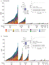
Figure 3.. Performance of the 10-gene marker panel in distinguishing between benign and cancer tissue.
FFPE samples of IDC/DCIS and Benign/Normal tissues were assayed by QM-MSP. Histogram plots indicate the percent methylation (colored segment) and cumulative methylation (bar height, Y-axis) for each of the 10-gene markers in the panel. Insets of 1) Box plots show the median cumulative methylation of cancer (IDC/DCIS) versus benign/normal tissues. 2) ROC analyses indicate the discriminatory power of the 10-gene marker panel. A. Training set. The laboratory threshold that provided highest sensitivity and specificity for detection of cancer in the training cohort was 14.5 CMI, based on ROC analysis. B. Test set. Conditions established in the training set were locked and performance of the test set was evaluated as in A. Insets of 1) Box plots show the median cumulative methylation of cancer (IDC/DCIS) versus benign/normal tissues. 2) ROC analyses indicate the discriminatory power of the 10-gene marker panel. The dashed line indicates our target sensitivity at 90%, and dot indicates the cutoff at 14.5 CMI units on the curve. CMI, cumulative methylation index; IDC, invasive ductal carcinoma; ILC, invasive lobular carcinoma, DCIS, ductal carcinoma in situ; FA, fibroadenoma; Pap, papilloma; UDH, usual ductal hyperplasia, and cysts. ROC, receiver operator characteristic; AUC, area under the ROC curve. Mann-Whitney P-values are indicated.
Figure 4.
Figure 4.. Assay validation- Interassay and interoperator reproducibility of the automated cartridge assay.
A. Box-whisker plots show the median and full range of cumulative methylation (CM) (Y-axis) for the 10-gene marker panel using fully methylated CAMA-1 DNA diluted with unmethylated human sperm DNA. The figure shows interassay reproducibility using five-six replicates for each sample (X-axis). Statistical significance is indicated by P values. B. Interoperator reproducibility was evaluated for 10 genes in a set of 23 patient FNA lysates analyzed independently by two investigators. CM measurements for the 10 markers between operators showed an intraclass coefficient (ICC) of 0.99. Kappa statistic for interrater reliability, using the threshold of 14.5 CMI from the training cohort, was 0.91 (95% CI: 0.75–1.08, p value: 6.76e-6).
Figure 5.
Figure 5.. Pilot test of the automated breast cancer detection cartridge on FNA samples.
Histogram shows the 10-gene marker panel distinguished between IDC (N=24) and benign (N=49) FNA samples from Portugal and Hong Kong, using a cutoff of CM 14.5 that was derived from training cohort of FFPE samples (denoted by horizontal line). Insets of 1) Box plot shows that cumulative methylation is significantly higher in IDC compared to benign breast FNA. 2) ROC analyses indicate the discriminatory power of the 10-gene marker panel. The dashed line indicates our target sensitivity at 90%, and dot indicates the cutoff at 14.5 CMI units on the curve. CM, cumulative methylation, IDC: Invasive ductal carcinoma, FNA: fine needle aspiration, N= number of samples.

References

    1. Jemal A, Bray F, Center MM, Ferlay J, Ward E, Forman D. Global cancer statistics. CA Cancer J Clin. 2011;61:69–90. - PubMed
    1. Soerjomataram I, Lortet-Tieulent J, Parkin DM, Ferlay J, Mathers C, Forman D, et al. Global burden of cancer in 2008: a systematic analysis of disability-adjusted life-years in 12 world regions. Lancet. 2012;380:1840–50. - PubMed
    1. Forouzanfar MH, Foreman KJ, Delossantos AM, Lozano R, Lopez AD, Murray CJ, et al. Breast and cervical cancer in 187 countries between 1980 and 2010: a systematic analysis. Lancet. 2011;378:1461–84. - PubMed
    1. Shulman LN, Willett W, Sievers A, Knaul FM. Breast cancer in developing countries: opportunities for improved survival. J Oncol. 2010;2010:595167. - PMC - PubMed
    1. Ezzati M, Pearson-Stuttard J, Bennett JE, Mathers CD. Acting on non-communicable diseases in low- and middle-income tropical countries. Nature. 2018;559:507–16. - PubMed

Publication types

MeSH terms

Substances