. 2019 Aug 21;39(34):6656-6667.
doi: 10.1523/JNEUROSCI.0278-19.2019. Epub 2019 Jul 12.

Axonal Growth of Midbrain Dopamine Neurons is Modulated by the Cell Adhesion Molecule ALCAM Through Trans-Heterophilic Interactions with L1cam, Chl1, and Semaphorins

Affiliations

Axonal Growth of Midbrain Dopamine Neurons is Modulated by the Cell Adhesion Molecule ALCAM Through Trans-Heterophilic Interactions with L1cam, Chl1, and Semaphorins

Christopher R Bye et al. J Neurosci. .

Abstract

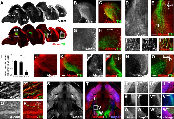
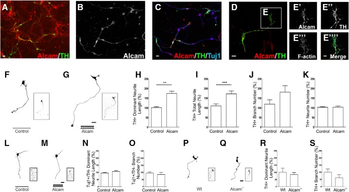
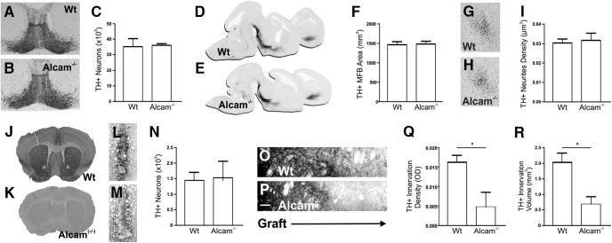
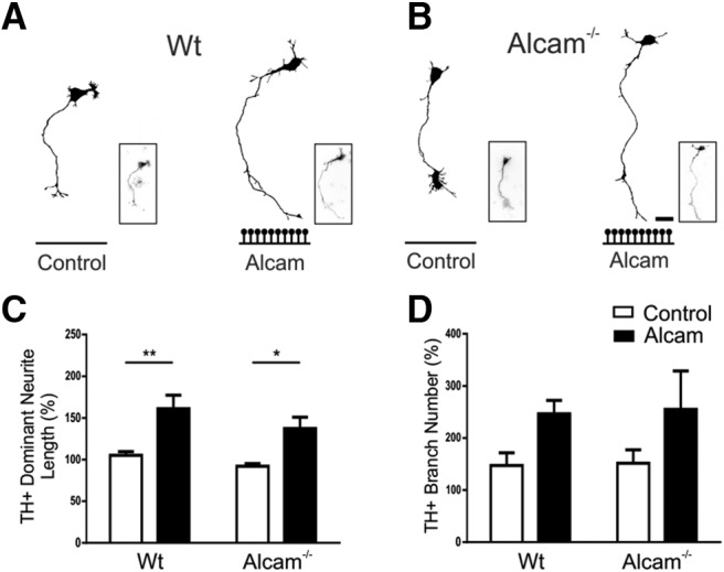
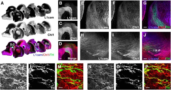
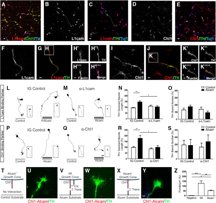
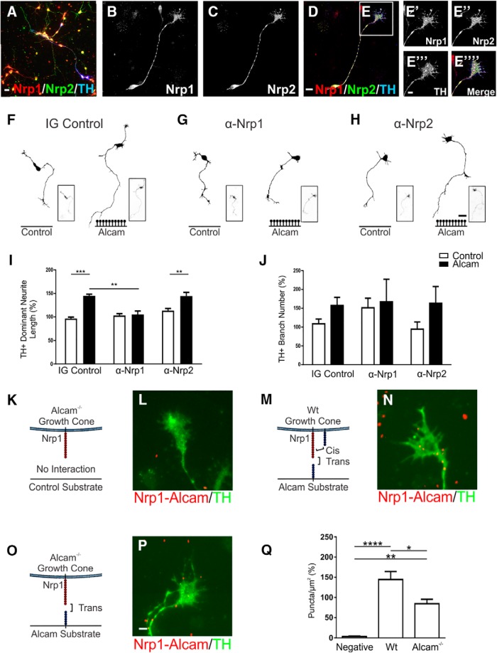
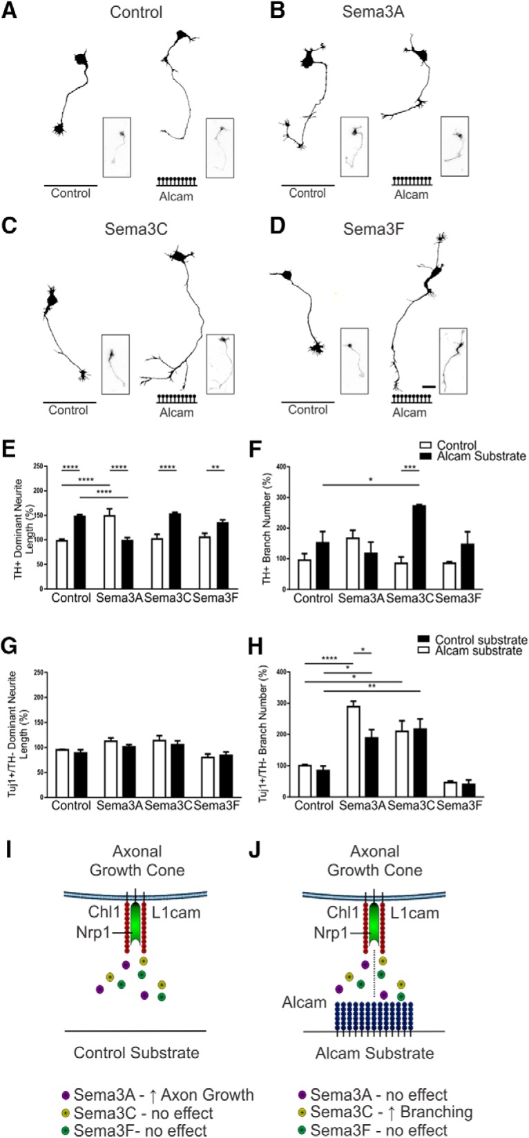
The growth of axons corresponding to different neuronal subtypes is governed by unique expression profiles of molecules on the growth cone. These molecules respond to extracellular cues either locally though cell adhesion interactions or over long distances through diffusible gradients. Here, we report that that the cell adhesion molecule ALCAM (CD166) can act as an extracellular substrate to selectively promote the growth of murine midbrain dopamine (mDA) neuron axons through a trans-heterophilic interaction with mDA-bound adhesion molecules. In mixed-sex primary midbrain cultures, the growth-promoting effect of ALCAM was abolished by neutralizing antibodies for components of the Semaphorin receptor complex Nrp1, Chl1, or L1cam. The ALCAM substrate was also found to modulate the response of mDA neurites to soluble semaphorins in a context-specific manner by abolishing the growth-promoting effect of Sema3A but inducing a branching response in the presence of Sema3C. These findings identify a previously unrecognized guidance mechanism whereby cell adhesion molecules act in trans to modulate the response of axonal growth cones to soluble gradients to selectively orchestrate the growth and guidance of mDA neurons.SIGNIFICANCE STATEMENT The mechanisms governing the axonal connectivity of midbrain dopamine (mDA) neurons during neural development have remained rather poorly understood relative to other model systems for axonal growth and guidance. Here, we report a series of novel interactions between proteins previously not identified in the context of mDA neuronal growth. Significantly, the results suggest a previously unrecognized mechanism involving the convergence in signaling between local, adhesion and long-distance, soluble cues. A better understanding of the molecules and mechanisms involved in establishment of the mDA system is important as a part of ongoing efforts to understand the consequence of conditions that may result from aberrant connectivity and also for cell replacement strategies for Parkinson's disease.

Keywords: CAMs; Nrp1; connectivity; guidance; mesencephalon.


Figures

Figure 1.
Figure 1.
Alcam in vivo expression during formation of the midbrain dopaminergic axonal pathways. In vivo representative sagittal images of whole embryonic brain at E12.5 (A) immunolabeled for Alcam and Alcam/TH showing Alcam expression relative to the developing dopaminergic pathway (displayed in a medial to lateral series). Inserts show high Alcam intensity in the ventral midbrain (B,C), including in dopamine neurons, and along the axonal pathway (D,E). Magnified high-resolution images show the expression of Alcam in both dopaminergic and nondopaminergic axons and cells along the pathway (F). Filled arrows indicate Alcam as a substrate expressed along the pathway and empty arrows indicate Alcam expression in TH+ axons. In the forebrain (G,H), lower Alcam intensity was observed in the GE, with higher intensity observed in nondopaminergic fibers at the boundary of the GE and cortex. Quantification of Alcam gene expression by qRT-PCR (I) showed a significant difference (ANOVA, F(2,7) = 17.02, p = 0.0021), with reduced expression at E14.5 compared with E10.5 (ANOVA, t = 5.263, df = 7, p = 0.0029, n = 4) and E12.5 levels (ANOVA, t = 4.528, df = 7, p = 0.0066, n = 4). At E14.5, Alcam protein expression was restricted to a caudomedial subpopulation of dopaminergic neurons in the ventral midbrain (J,K) and largely absent in dopaminergic and nondopaminergic axons along the pathway (L,M) and in the GE (N,O). Strong Alcam intensity continued in nondopaminergic fibers at the boundary of the GE and cortex, magnified images show mesocortical dopaminergic axons wrapping around strongly expressing Alcam fibers descending from the developing cortex into the GE (P). Dopaminergic axons were also observed wrapping around intensely Alcam-labeled fibers in the mesohabenular pathway exiting the midbrain (Q,R), schematic of image location shown at top right. Horizontal image of whole adult brain (S,T) immunolabeled for Alcam/TH/Darp32 showing no Alcam expression in the fully formed mDA pathway. Inserts show Alcam was expressed intensely in Darp32+ medium spiny neurons and axons, not in dopaminergic neurons and axons in the striatum (U), along the dopaminergic axonal pathways (V), and in the ventral midbrain (W). Scale bars: BE, 20 μm; F, 10 μm; G, H, JO, U, W, 20 μm. Data are shown as mean ± SEM. Ctx; Cortex; FB, forebrain; GE, ganglionic eminence; HB, hindbrain; Hyp, hypothalamus; MB, midbrain. **p < 0.01.
Figure 2.
Figure 2.
Alcam in vitro expression and function during dopaminergic axonal growth. Representative images of neurons in primary midbrain cultures immunolabeled for Alcam (AC) show Alcam expression in dopaminergic and nondopaminergic neurons. Nondopaminergic neuronal populations display a range of expression from very strong, to completely absent. In dopaminergic neurons, punctate Alcam expression was present in cell bodies and axons (D) and in F-actin+ growth cones (E). Representative images and corresponding axonal traces of TH+ neurons in primary midbrain cultures grown on control (F), or high-density Alcam substrate (G). Quantification of axon growth in the presence of an Alcam substrate resulted in a significant increase in length of the longest (dominant) neurite (H) (unpaired t test, t = 4.533, df = 8, p = 0.0019, n = 5) and the length of all (total) neurites per neuron (I) (unpaired t test, t = 2.659, df = 8, p = 0.0289, n = 5). No significant effect was observed on number of branches (J) or number of neurites (K) per neuron. Representative traces of nondopaminergic midbrain neuron traces (Tuj1+/TH) from primary midbrain neurons cultured on control (L) or high-density Alcam substrate (M). Quantification of axon growth in the presence of Alcam substrate resulted in no significant effect on neurite length (N) or branch number (O) (n = 5). Representative axonal traces of dopaminergic (TH+) neurons in primary midbrain cultures from WT (P) and Alcam−/− mice (Q). Quantification of axon growth showed no significant changes due to loss of Alcam function on (R) neurite length or (S) branch number (unpaired t test, n = 4). Scale bars: AD, 20 μm; E, 10 μm; F, G, L, M, P, Q, 20 μm. Data are shown as mean ± SEM. **p < 0.01, ***p < 0.001.
Figure 3.
Figure 3.
Alcam loss of function in midbrain dopamine neurons during axonal pathway formation in vivo. Representative coronal images of the midbrain at E14.5 in WT (A) and Alcam−/− (B) mice showed no significant difference in immunolabeled TH neuron number (C) (unpaired t test, n = 4). Sagittal images of the developing midbrain dopamine axonal pathway at E14.5 in WT (D) and Alcam−/− (E) mice immunolabeled for TH revealed no gross defect in pathway growth or trajectory. Quantification of total volume (F) of the axon bundle, or in the density of dopaminergic varicosities (GI) reaching the forebrain revealed no significant difference in axon fasciculation or pathfinding (unpaired t test, n = 4). Coronal images of adult mouse forebrain immunolabeled for Alcam show extensive expression throughout the striatum of WT mice (J), but completely absent in Alcam−/− mice (K). Six weeks after transplantation of dissociated E12.5 ventral midbrain neurons into the striatum of WT (L) or Alcam−/− mice (M), coronal images show no significant difference in TH immunolabeled neuron survival (N) (unpaired t test, n = 4). Magnified images immediately adjacent to the transplanted neuron graft (O,P) show a significant reduction of 0.0113 ± 0.0048 in the density of TH innervation (Q) (unpaired t test, t = 2.340, df = 8, p = 0.0474, n = 4) in the Alcam−/− host. Quantification of axonal innervation volume from WT neurons into an Alcam−/− host resulted in a significant reduction of 1.342 ± 0.3782 mm3 in the volume of innervation (R) (unpaired t test, t = 2.538 df = 8, p = 0.0075, n = 4). Scale bars in O and P, 10 μm. Data are shown as mean ± SEM. *p < 0.05.
Figure 4.
Figure 4.
Functional analysis of Alcam homophilic and trans-heterophilic interactions in the promotion of axon growth by an Alcam substrate. Representative TH+ axonal traces from primary ventral midbrain cultures of WT (A) or Alcam−/− (B) neurons cultured on control or Alcam substrate. Quantification of axon growth in the presence of an Alcam substrate resulted in a significant increase in neurite length (C) (two-way ANOVA, Alcam substrate, F(1,12) = 23.05, p = 0.004, n = 4) in both WT (t = 3.741, df = 12, p = 0.0056) and Alcam−/− neurons (t = 3.049, df = 12, p = 0.0201), where loss of Alcam function on growing axons prevents homophilic interactions. A significant effect on branch number (D) (two-way ANOVA, substrate, F(1,12) = 5.554, p = 0.0363, n = 4) was not significant in post hoc tests. Scale bars in A and B, 20 μm. Data are shown as mean ± SEM. *p < 0.05, **p < 0.01.
Figure 5.
Figure 5.
Expression of L1cam and Chl1 in vivo during formation of the midbrain dopaminergic axonal pathways. Sagittal images of whole embryonic brain at E12.5 (A) immunolabeled for L1cam, Chl1, and L1cam/Chl1/TH showing their expression in the developing dopaminergic pathway (displayed in a medial to lateral series). Inserts show both L1cam and Chl1 were highly expressed in the ventral midbrain (BD), including in dopaminergic neurons along the mDA axonal pathway (EG) in both dopaminergic and nondopaminergic axons. In the forebrain (HJ), L1cam and Chl1 expression was located in both dopaminergic axons and nondopaminergic axons, with prominent expression in the cortex. Magnified images show colocalization maps of L1cam (KM) or Chl1 (NP) in dopaminergic axons and growth cones at the axonal front. Scale bars: BJ, 20 μm; KP, 10 μm. BS, Brainstem; Ctx, cortex; FB, forebrain; GE, ganglionic eminence; MB, midbrain.
Figure 6.
Figure 6.
Functional analysis of Alcam trans-heterophilic interactions with the potential binding partners L1cam and Chl1. Representative images of primary midbrain cultures immunolabeled for the potential Alcam trans-heterophilic binding partners L1cam and Chl1 show broad expression of both CAM's in dopaminergic and nondopaminergic neurons (A). Expression was localized throughout the cell body, axons, and branches of all neurons for L1cam (B,C), and Chl1 (D,E). In dopaminergic neurons, L1cam expression was evenly distributed in cell bodies, axons (F,G), and F-actin+ growth cones (H). Parallel images of the potential Alcam-binding partner Chl1 (I,J) also show Chl1 expression in cell bodies, axons, and F-actin+ growth cones (K). Representative dopaminergic (TH+) neuron traces of WT neurons cultured on control or Alcam substrate in the presence of an IG control (L) or L1cam-neutralizing antibody (M), blocking potential trans heterophilic interactions between Alcam and L1cam. A significant interaction (two-way ANOVA, F(1,8) = 9.264, p = 0.0160) blocking antibody (F(1,8) = 6.531, p = 0.0339) and substrate (F(1,8) = 17.18, p = 0.0032) effect was detected (n = 3). The significant growth promoting effect of Alcam substrate on neurite length (N) (t = 5.083, df = 8, p = 0.0019) was ablated in the presence of functional blocking of L1cam (t = 0.7783, df = 8, p = 0.7071). Neurite length decreased significantly in a direct comparison of neurons grown on Alcam substrate in control and in the presence of the L1cam blocking antibody (t = 3.959, df = 8, p = 0.0248). No significant effect on branch number (O) was observed. Representative dopaminergic neuron traces of WT neurons cultured on control or Alcam substrate in the presence of IG control (P) or Chl1-neutralizing antibody (Q), blocking potential trans heterophilic interactions between Alcam and Chl1. A significant interaction (two-way ANOVA, F(1,8) = 5.650, p = 0.0447) blocking antibody (F(1,8) = 10.52, p = 0.0118) and substrate (F(1,8) = 9.549, p = 0.0149) effect was detected (n = 3). The significant growth promoting effect of Alcam substrate on neurite length (R) (t = 3.866, df = 8, p = 0.0095) was ablated in the presence of functional blocking of Chl1 (t = 0.5042, df = 8, p = 0.8614). Neurite length decreased significantly in a direct comparison of neurons grown on Alcam substrate in control and in the presence of the Chl1 blocking antibody (t = 3.975, df = 8, p = 0.0243). No significant effect on branch number (S) was observed. Representative immunohistochemistry images of primary dopaminergic growth cones immunolabeled using an in situ proximity ligation assay to detect interactions between Alcam and Chl1. Puncta (shown in red) indicate positive Alcam-Chl1 interactions in Alcam−/− neurons grown on a control substrate (T, U, no cis or trans interactions), WT neurons cultured on Alcam substrate (V, W, cis and trans interactions) and Alcam−/− neurons grown on an Alcam substrate (X, Y, trans interactions only). Quantification of Alcam-Chl1+ puncta (Z) showed a significant difference between groups (one-way ANOVA, F(2,8) = 27.00, p = 0.0003, n = 3–4). Relative to the negative control group, a significant increase in puncta was observed in WT neurons grown on Alcam substrate (t = 10.38, df = 8, p = 0.0002) and in Alcam−/− neurons grown on Alcam substrate (t = 4.420 df = 8, p = 0.0338). Comparison of WT and Alcam−/− neurons on Alcam substrate resulted in a significant 40% reduction in puncta (t = 46.291, df = 8, p = 0.0054) attributable directly to Alcam-Chl1 trans heterophilic interactions. Scale bars: AE, 20 μm; F, G, 10 μm; H, 5 μm; I, J, 10 μm; K, 5 μm; L, M, P, Q, 20 μm; U, W, Y, 5 μm. Data are shown as mean ± SEM. *p < 0.05, **p < 0.01, ***p < 0.001.
Figure 7.
Figure 7.
Functional analysis of Alcam trans-heterophilic interactions with the Semaphorin receptors Nrp1 and Nrp2. Representative images of primary midbrain cultures immunolabeled for the potential Alcam trans-heterophilic binding partners Nrp1 and Nrp2 show broad expression of both receptors in dopaminergic and nondopaminergic neurons (A). In dopaminergic neurons, Nrp1 and Nrp2 expression was evenly distributed in cell bodies, axons (BD) and F-actin+ growth cones (E). Representative dopaminergic (TH+) neuron traces of WT primary midbrain neurons cultured on control or Alcam substrate in the presence of IG control (F), Nrp1 (G), or Nrp2 (H)-neutralizing antibody, blocking potential trans-heterophilic interactions between Alcam and Nrp1 or Nrp2. Neurite length (I) of dopaminergic neurons showed a significant interaction (two-way ANOVA, F(2,12) = 9.385, p = 0.0035) blocking antibody (F(2,12) = 10.87, p = 0.0020) and substrate (F(1,12) = 38.29, p < 0.0001) effect (n = 3). The significant growth promoting effect of Alcam substrate on neurite length (t = 6.354, df = 12, p = 0.0001) was ablated in the presence of functional blocking of the Nrp1 (t = 0.2888, df = 12, p = 0.9890), but not Nrp2 receptor (t = 4.076, df = 12, p = 0.0046). No significant effect on (J) branch number was observed (two-way ANOVA, n = 3). Representative immunohistochemistry images of primary dopaminergic growth cones immunolabeled using an in situ proximity ligation assay to detect interactions between Alcam and Nrp1. Puncta (shown in red) indicating positive Alcam-Nrp1 interactions in Alcam−/− neurons grown on a control substrate (K, L, no cis or trans interactions), WT neurons cultured on Alcam substrate (M, N, cis and trans interactions) and Alcam−/− neurons grown on an Alcam substrate (O, P, trans interactions only). Quantification of Alcam-Nrp1+ puncta (Q) showed a significant difference between groups (one-way ANOVA, F(2,9) = 29.10, p = 0.0001, n = 4). Relative to the negative control group, a significant increase in puncta was observed in WT neurons grown on Alcam substrate (t = 10.75, df = 9, p < 0.0001) and in Alcam−/− neurons grown on Alcam substrate (t = 6.172, df = 9, p = 0.0046). Comparison of WT and Alcam−/− neurons on Alcam substrate resulted in a significant 58% reduction in puncta (t = 4.577, df = 9, p = 0.0250) attributable directly to trans heterophilic interactions Scale bars: A, 20 μm; BD, 10 μm; E, 5 μm; FH, 20 μm; L, N, P, 5 μm. Data are shown as mean ± SEM. *p < 0.05, **p < 0.01, ***p < 0.001, ****p < 0.0001.
Figure 8.
Figure 8.
Functional analysis of Alcam substrate modulation of Semaphorin signaling. Representative dopaminergic (TH+) neuron traces of WT primary midbrain neurons cultured on control or Alcam substrate and treated with control (A), Sema3A (B), Sema3C (C), or Sema3F (D). Neurite length (E) of dopaminergic neurons showed a significant interaction (two-way ANOVA, F(3,16) = 28.94, p < 0.0001) and substrate (F(1,16) = 19.59, p = 0.0004) effect (n = 3). When grown on Alcam substrate, neurite length was significantly increased in control (t = 5.580, df = 16, p = 0.0002), significantly decreased with Sema3A treatment (t = 5.677, df = 16, p = 0.0001), significantly increased with Sema3C treatment (t = 5.697, df = 16, p = 0.0001), and significantly increased with Sema3F treatment (t = 3.253, df = 16, p = 0.0198). Direct comparison of Semaphorin-treated neurite length on control substrate significantly increased only with Sema3A treatment (t = 5.729, df = 16, p = 0.0009). The significant growth promoting effect of Sema3A and Alcam substrate on neurite length was reversed in the presence of both Sema3A and Alcam substrate (t = 5.529, df = 16, p < 0.0013). Branch number (F) of dopaminergic neurons showed a significant interaction (two-way ANOVA, F(3,16) = 6.166, p = 0.0055) and substrate (F(1,16) = 10.80, p = 0.0046) effect (n = 3). Branch number significantly increased only in dopaminergic neurons grown on an Alcam substrate and treated with Sema3C (t = 4.808, df = 16, p = 0.0008). Branch number increased significantly in a direct comparison of control and Sema3A-treated neurons grown on Alcam substrate (t = 3.093, df = 16, p = 0.0300), with no significant effect observed in control and Sema3A or Sema3F treated cultures. In nondopaminergic neurons (TH/Tuj1+), a significant effect on neurite length due to Alcam substrate following Semaphorin treatment (G) (two-way ANOVA, Sema treatment, F(3,16) = 7.213, p = 0.0028, n = 3) was not significant in post hoc tests. A significant effect on branch number due to Alcam substrate following Semaphorin treatment (H) (two-way ANOVA, Sema treatment, F(3,16) = 37.66, p < 0.0001, n = 3) was also observed. Sema3A treatment resulted in a significant decrease in branch number (H) in nondopaminergic neurons when grown on an Alcam substrate (t = 3.260, df = 16, p = 0.0195). Branch number increased significantly in a direct comparison of control and Sema3A-treated neurons grown on control substrate (t = 6.142, df = 16, p = 0.0004), but not in Alcam substrate (t = 3.404, df = 16, p = 0.0968). Schematic of the regulation of Semaphorin signaling the absence (I) or presence (J) of Alcam substrate indicating the proposed trans-heterophilic interactions with the semaphorin receptor complex (comprised of Nrp1, L1cam and Chl1) on the growth cone. Scale bars in AD, 20 μm. Data are shown as mean ± SEM. *p < 0.05, **p < 0.01, ***p < 0.001, ****p < 0.0001.

Similar articles

Cited by

References

    1. Alsanie WF, Penna V, Schachner M, Thompson LH, Parish CL (2017) Homophilic binding of the neural cell adhesion molecule CHL1 regulates development of ventral midbrain dopaminergic pathways. Sci Rep 7:9368. 10.1038/s41598-017-09599-y - DOI - PMC - PubMed
    1. Andrews WD, Barber M, Nemitz M, Memi F, Parnavelas JG (2017) Semaphorin3A-neuropilin1 signalling is involved in the generation of cortical interneurons. Brain Struct Funct 222:2217–2233. 10.1007/s00429-016-1337-3 - DOI - PMC - PubMed
    1. Avci HX, Zelina P, Thelen K, Pollerberg GE (2004) Role of cell adhesion molecule DM-GRASP in growth and orientation of retinal ganglion cell axons. Dev Biol 271:291–305. 10.1016/j.ydbio.2004.03.035 - DOI - PubMed
    1. Brignani S, Pasterkamp RJ (2017) Neuronal subset-specific migration and axonal wiring mechanisms in the developing midbrain dopamine system. Front Neuroanat 11:55. 10.3389/fnana.2017.00055 - DOI - PMC - PubMed
    1. Blakely BD, Bye CR, Fernando CV, Horne MK, Macheda ML, Stacker SA, Arenas E, Parish CL (2011) Wnt5a regulates midbrain dopaminergic axon growth and guidance. PloS one 6:e18373. 10.1371/journal.pone.0018373 - DOI - PMC - PubMed

Publication types