. 2019 Jul 30;116(31):15635-15644.
doi: 10.1073/pnas.1904714116. Epub 2019 Jul 12.

CRISPR-Cas9-mediated gene knockout in intestinal tumor organoids provides functional validation for colorectal cancer driver genes

Affiliations

CRISPR-Cas9-mediated gene knockout in intestinal tumor organoids provides functional validation for colorectal cancer driver genes

Haruna Takeda et al. Proc Natl Acad Sci U S A. .

Abstract

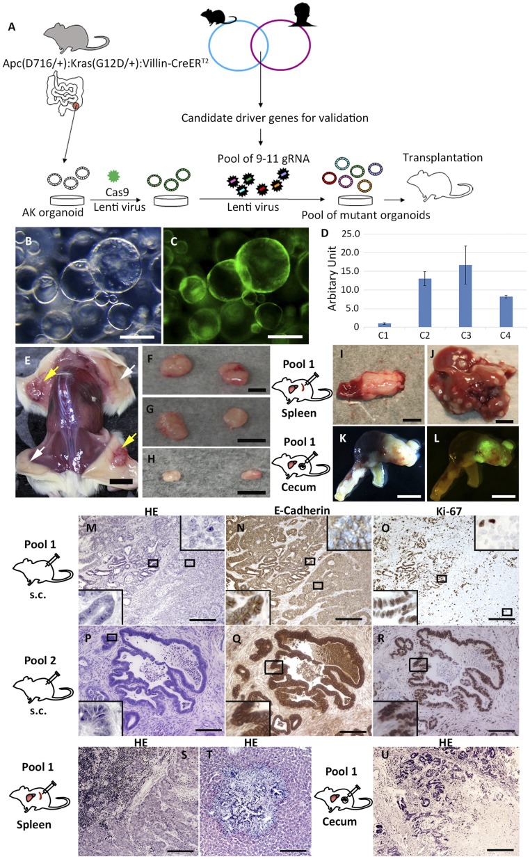
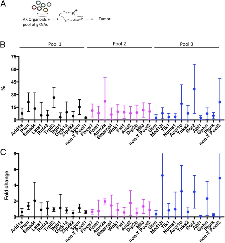
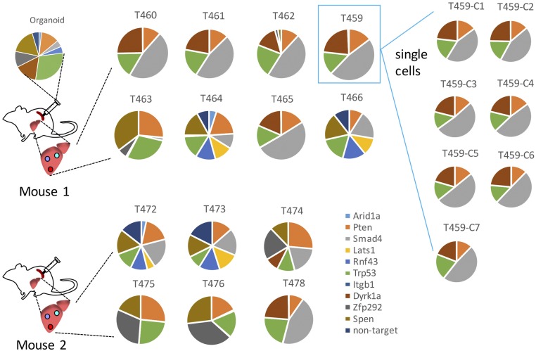
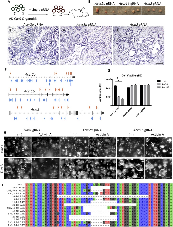
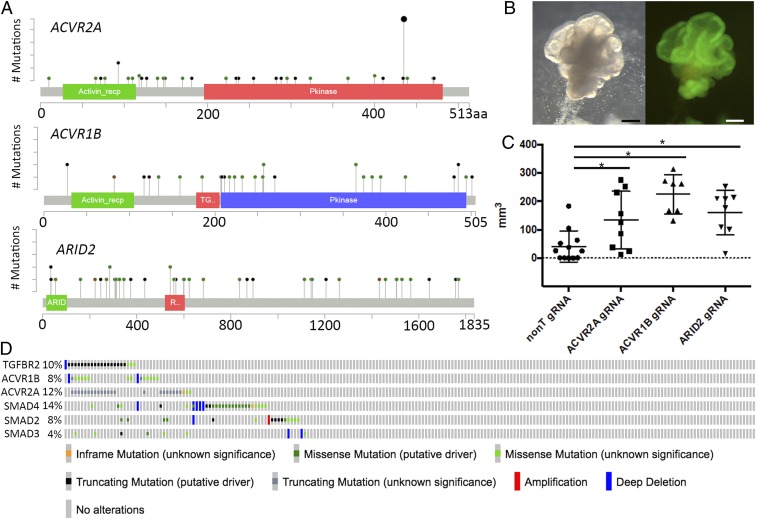
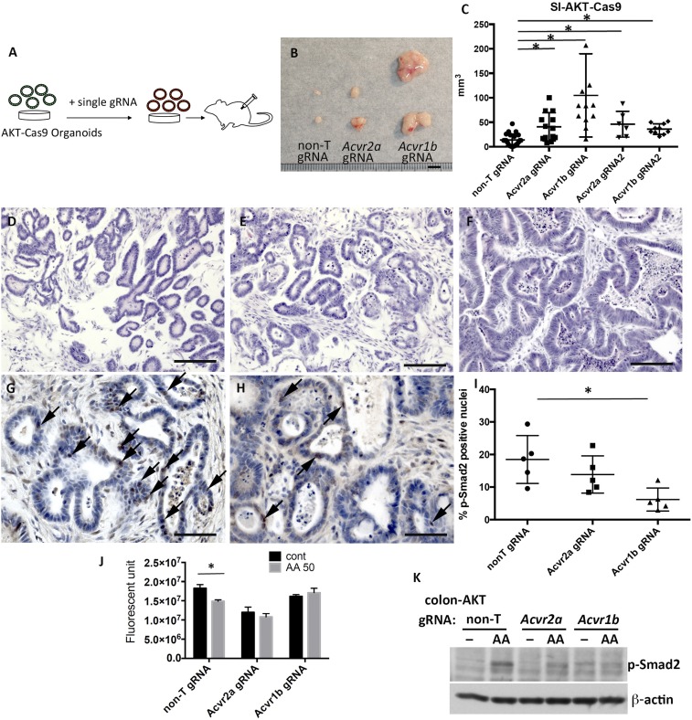
Colorectal cancer (CRC) is the third leading cause of cancer-related deaths worldwide. Several genome sequencing studies have provided comprehensive CRC genomic datasets. Likewise, in our previous study, we performed genome-wide Sleeping Beauty transposon-based mutagenesis screening in mice and provided comprehensive datasets of candidate CRC driver genes. However, functional validation for most candidate CRC driver genes, which were commonly identified from both human and mice, has not been performed. Here, we describe a platform for functionally validating CRC driver genes that utilizes CRISPR-Cas9 in mouse intestinal tumor organoids and human CRC-derived organoids in xenograft mouse models. We used genetically defined benign tumor-derived organoids carrying 2 frequent gene mutations (Apc and Kras mutations), which act in the early stage of CRC development, so that we could clearly evaluate the tumorigenic ability of the mutation in a single gene. These studies showed that Acvr1b, Acvr2a, and Arid2 could function as tumor suppressor genes (TSGs) in CRC and uncovered a role for Trp53 in tumor metastasis. We also showed that co-occurrent mutations in receptors for activin and transforming growth factor-β (TGF-β) synergistically promote tumorigenesis, and shed light on the role of activin receptors in CRC. This experimental system can also be applied to mouse intestinal organoids carrying other sensitizing mutations as well as organoids derived from other organs, which could further contribute to identification of novel cancer driver genes and new drug targets.

Keywords: CRISPR-Cas9; activin; colorectal cancer; driver gene; organoid.


Conflict of interest statement

The authors declare no conflict of interest.

Figures

Fig. 1.
Fig. 1.
Candidate colorectal driver gene validation using mouse intestinal tumor organoids and CRISPR-Cas9. (A) Schematic view of the experimental system. Mouse intestinal tumor organoids were established from ApcΔ716:Kras+/G12D:Villin-CreERT2 mice (19). A Cas9 lentivirus containing a GFP expression cassette was introduced into the intestinal organoids. Pools of gRNAs targeting 9 to 10 unique candidate TSGs were then introduced into the GFP(+) organoids and subsequently transplanted into mice and monitored for tumor formation. (B) Macroscopic view of Cas9-expressing AK organoids. (C) GFP-positive organoids indicate that the plasmid encoding Cas9 was introduced into the organoids. (D) qPCR analysis for Cas9 messenger RNA showed that C2 and C3 organoids showed more than 10-fold higher expression compared with C1 organoids. (E and F) Macroscopic views of s.c. tumors derived from organoids with pool 1. Yellow arrows indicate s.c. tumors derived from organoids with pool 1. White arrows indicate where AK-Cas9-gRNA (nontarget) organoids were transplanted. (G) s.c. tumors derived from organoids with pool 2. (H) s.c. tumors derived from organoids with pool 3. A spleen tumor (I) and liver tumors (J), both derived from organoids with pool 1, are shown. (K) Bright-field photograph of a primary cecum tumor derived from organoids with pool 1. (L) Tumor cells derived from organoids were GFP-positive. Hematoxylin/eosin (HE) staining of s.c. tumors derived from organoids with pool 1 (M) and pool 2 (P) is shown. Immunohistochemistry (IHC) for E-cadherin on a s.c. tumor derived from organoids with pool 1 (N) and pool 2 (Q) is shown. IHC for Ki-67 on a s.c. tumor derived from organoids with pool 1 (O) and pool 2 (R) is shown. (MR, Insets) Photographs with higher magnification. HE staining of a splenic tumor (S) and a metastatic liver tumor (T) derived from organoids with pool 1 is shown. (U) HE staining of a primary cecum tumor derived from organoids with pool 1 in an orthotopic model. (Scale bars: B and C, 250 μm; E, 1 cm; FL, 5 mm; MU, 200 μm.)
Fig. 2.
Fig. 2.
Analysis of gRNA frequencies in tumors by next-generation sequencing. (A) Scheme describing the experimental flow. (B) Average fraction of each gRNA in tumor tissues. (C) Average fold change of each gRNA fraction in tumors compared with preinjected organoids.
Fig. 3.
Fig. 3.
Analysis of gRNA frequencies. The pie charts show the fraction of gRNA in organoids, liver tumors, and organoids derived from single cells of tumor T459.
Fig. 4.
Fig. 4.
Knockout of Acvr2a, Acvr1b, or Arid2 in AK organoids promotes s.c. tumor development. (A) Method used to test the ability of single Acvr2a, Acvr1b, or Arid2 knockouts in AK organoids to induce s.c. tumors following transplantation to NSG mice. (B) Macroscopic appearance of s.c. tumors derived from AK organoids carrying Acvr2a gRNA (Left), Acvr1b gRNA (Center), and Arid2 gRNA (Right). Hematoxylin/eosin (HE) staining of s.c. tumors derived from AK organoids containing gRNA for Acvr2a (C), Acvr1b (D), or Arid2 (E) is shown. (F) SB transposon insertion patterns were obtained from a previous study (10) for Acvr2a, Acvr1b, and Arid2. Orange arrowheads represent transposon insertions with a forward orientation relative to the transcript. Blue arrowheads represent transposon insertions with a reverse orientation relative to the transcript. (G) Bar graph indicates cell viability in the absence of activin A or in the presence of activin A (50 ng/mL or 100 ng/mL). AA, activin A; cont, control. *P < 0.05 by t test. (H) Photographs show colonic AK organoids with gRNA for non-T, Acvr2a, or Acvr1b in the presence or absence of activin A (50 ng/mL) at day 1 (Upper) and day 3 (Lower). (I) Deep sequencing analysis of the CRISPR on-target site in the Acvr1b gene. The majority of sequence reads showed deletions of insertions that resulted in frameshift mutations. del, deletion; NS, nucleotide substitution. (Scale bars: B, 5 mm; CE, 200 μm; H, 100 μm.)
Fig. 5.
Fig. 5.
Analysis of ACVR2A, ACVR1B, and ARID2 mutations in human CRC. (A) Spectrum of mutations in ACVR2A, ACVR1B, and ARID2 in human CRC. The mutational results are based on the data generated by the The Cancer Genome Atlas Research Network study (5) (https://www.cancer.gov/about-nci/organization/ccg/research/structural-genomics/tcga). Black circles denote termination mutations, while green circles denote nonsynonymous mutations. (B) Transduction of Cas9 to human CRC-derived organoids. The plasmid contains both a Cas9 cDNA and a GFP cDNA; therefore, Cas9-expressing organoids can be visualized by GFP. (Left) Bright field. (Right) GFP. (C) Transduction of ARID2 gRNA or ACVR1B gRNA or ACVR2A into Cas9-expressing human CRC organoids promoted tumor growth compared with parental organoids. *P < 0.05 by t test. (D) Oncoprint showing the frequencies of mutations for genes belonging to the TGF-β superfamily in CRC. (Scale bars: 200 μm.)
Fig. 6.
Fig. 6.
Acvr2a or Acvr1b mutations synergistically cooperate with Tgfbr2 mutations in AKT organoids in tumor development. (A) Scheme describing the experimental design to knock out Acvr1b and Acvr2a in AKT organoids. (B) Macroscopic view of the s.c. tumors derived from AKT-Cas9 organoids carrying non-T gRNA, Acvr2a gRNA, or Acvr1b gRNA. (C) Graph shows the tumor size distribution for AKT-Cas9 organoids carrying non-T gRNA, Acvr2a gRNA, or Acvr1b gRNA. SI, small intestine. *P < 0.05 by χ2 test. Hematoxylin/eosin staining of s.c. tumors derived from AKT-Cas9 organoids carrying non-T gRNA (D), Acvr2a gRNA (E), or Acvr1b gRNA (F) is shown. Immunohistochemistry of p-Smad2 on the s.c. tumors derived from AKT-Cas9 non-T gRNA organoids (G) or AKT-Cas9 Acvr1b gRNA organoids (H) is shown. Black arrows indicate p-Smad2–positive nuclei in tumor epithelial cells. (I) Percentage of p-Smad2–positive nuclei in tumor epithelial cells. Five views were taken from 3 independent tumors, and >1,000 cells in total were counted. *P < 0.05. (J) Bar graph indicates cell viability in the absence of activin A or in the presence of activin A (AA; 50 ng/mL). cont, control. *P < 0.05 by t test. (K) Twelve hours after activin A (50 ng/mL) treatment, the level of p-Smad2 was analyzed by Western blotting. *P < 0.05 by t test. (Scale bars: B, 5 mm; DF, 100 μm; G and H, 50 μm.)

Similar articles

Cited by

References

    1. Kinzler K. W., Vogelstein B., Lessons from hereditary colorectal cancer. Cell 87, 159–170 (1996). - PubMed
    1. Pinto D., Clevers H., Wnt, stem cells and cancer in the intestine. Biol. Cell 97, 185–196 (2005). - PubMed
    1. Bos J. L., ras oncogenes in human cancer: A review. Cancer Res. 49, 4682–4689 (1989). - PubMed
    1. Baker S. J., et al. , p53 gene mutations occur in combination with 17p allelic deletions as late events in colorectal tumorigenesis. Cancer Res. 50, 7717–7722 (1990). - PubMed
    1. Cancer Genome Atlas Network , Comprehensive molecular characterization of human colon and rectal cancer. Nature 487, 330–337 (2012). - PMC - PubMed

Publication types

Substances