. 2019 Jul 30;116(31):15560-15569.
doi: 10.1073/pnas.1904684116. Epub 2019 Jul 12.

Cardiomyocyte orientation modulated by the Numb family proteins-N-cadherin axis is essential for ventricular wall morphogenesis

Affiliations

Cardiomyocyte orientation modulated by the Numb family proteins-N-cadherin axis is essential for ventricular wall morphogenesis

Lianjie Miao et al. Proc Natl Acad Sci U S A. .

Abstract

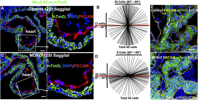
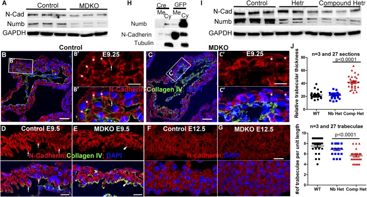
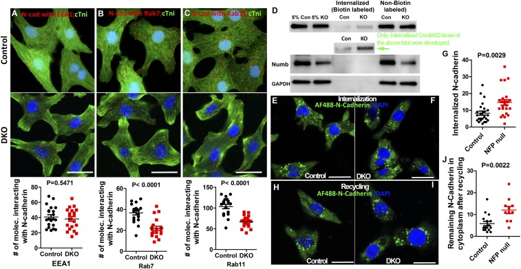
The roles of cellular orientation during trabecular and ventricular wall morphogenesis are unknown, and so are the underlying mechanisms that regulate cellular orientation. Myocardial-specific Numb and Numblike double-knockout (MDKO) hearts display a variety of defects, including in cellular orientation, patterns of mitotic spindle orientation, trabeculation, and ventricular compaction. Furthermore, Numb- and Numblike-null cardiomyocytes exhibit cellular behaviors distinct from those of control cells during trabecular morphogenesis based on single-cell lineage tracing. We investigated how Numb regulates cellular orientation and behaviors and determined that N-cadherin levels and membrane localization are reduced in MDKO hearts. To determine how Numb regulates N-cadherin membrane localization, we generated an mCherry:Numb knockin line and found that Numb localized to diverse endocytic organelles but mainly to the recycling endosome. Consistent with this localization, cardiomyocytes in MDKO did not display defects in N-cadherin internalization but rather in postendocytic recycling to the plasma membrane. Furthermore, N-cadherin overexpression via a mosaic model partially rescued the defects in cellular orientation and trabeculation of MDKO hearts. Our study unravels a phenomenon that cardiomyocytes display spatiotemporal cellular orientation during ventricular wall morphogenesis, and its disruption leads to abnormal trabecular and ventricular wall morphogenesis. Furthermore, we established a mechanism by which Numb modulates cellular orientation and consequently trabecular and ventricular wall morphogenesis by regulating N-cadherin recycling to the plasma membrane.

Keywords: Numb family proteins; cellular orientation; endocytosis; single-cell lineage tracing; trabecular morphogenesis.


Conflict of interest statement

The authors declare no conflict of interest.

Figures

Fig. 1.
Fig. 1.
NFPs are required for cellular orientation and organization during cardiac morphogenesis. Most of cardiomyocytes in the inner layer of the compact zone from a control heart orient perpendicularly to the heart wall (A and B), while most of the cells in the MDKO display no or parallel orientation to the heart wall (C and D). The double-headed arrows indicate the orientation of a cell and the asterisk indicates no orientation of a cell in A and C. B shows the cellular orientation of 49 cells of control hearts and D shows 50 cells in MDKO hearts. Each line represents an orientation of a cell in the inner layer in A or C and the red line represents the heart surface reference line in B and D. (E and F) The cells in control trabeculae display a long spindle shape and align parallel to trabecula (E), while cells in MDKO trabeculae do not show orientation and are round, indicated by an asterisk (F). (Scale bars in A, C, E, and F: 20 μm.)
Fig. 2.
Fig. 2.
NFPs regulate oriented cell division (OCD) and directional migration. Cardiomyocytes of single-layer myocardium undergo perpendicular OCD in A and parallel division in B. The mitotic spindles are identified by acetylated α-tubulin and endocardial cells are identified by endomucin. (C) The angle between mitotic spindles and the myocardial plain was measured and compared between control and MDKO at about E9.25. The patterns of mitotic spindle orientation between control and MDKO are different based on χ2 analysis. (D) Via single-cell lineage tracing, the GFP-labeled iDKO clone, indicated by the white arrow, does not express Numb based on ISH. (E) The clonal patterns between control and iDKO are significantly different, and the iDKO clones contain smaller percentages of trabecular and transmural clones and higher percentage of surface clones. (F and G) The MDKO hearts display a smaller number of trabeculae and the trabeculae in MDKO are shorter than control trabeculae. White arrows indicate trabeculae. (Scale bars in A, B, and D: 20 μm and in F and G: 50 μm.)
Fig. 3.
Fig. 3.
NFPs regulate transmural growth and cardiomyocyte proliferation autonomously. (A) Control hearts and (B) iDKO hearts show sporadic clones labeled with different colors. Cardiomyocytes in hearts were induced to be labeled at E7.75 with dosage of tamoxifen at 20 μg/g body weight and the hearts at E12.5 were imaged via Leica M205 FA, which can detect fluorescent protein-labeled cells at single-cell resolution. (CE) Individual clones in the cleared hearts were 3D-imaged to identify the geometric pattern of each of the clones. C, D, and E show compact, trabecular and transmural clones, respectively. (F) The clonal patterns between control and iDKO. (G) The transmural growth of the clones and that the iDKO clones grow significantly shorter distances compared with the control clones. (H) The number of cells in each clone; the iDKO clones contain significantly more cells than the control clones across all 3 types of clones. (Scale bars in A and B: 200 μm and in CE: 50 μm.)
Fig. 4.
Fig. 4.
NFPs regulate trabecular morphogenesis through N-cadherin. (A) MDKO hearts display lower levels of Numb and N-cadherin than control hearts at E13.5. (B) N-cadherin localizes to the lateral domain of the cardiomyocytes in the myocardium at E9.25, indicted by the asterisk, and to the lateral and apical domains of some cardiomyocytes that face toward the heart lumen, indicated by the arrow. (C) N-cadherin localization to the lateral domain is weaker in the MDKO, as indicated by the asterisk. (D and E) N-cadherin localizes to the apical domain strongly in the control heart at E9.5 but weakly in the MDKO heart. The weaker membrane localization in the MDKO is further demonstrated in E12.5 hearts. (F and G) The membrane fractionation shows that the membrane N-cadherin in cultured NFP null cardiomyocytes is significantly reduced compared with the controls (H). (I) Levels of Numb and N-cadherin proteins are reduced in the compound heterozygotes. (J) Compound heterozygous hearts (Nkx2.5Cre/+;Nbfl/fl; Nlfl/+;Cdh2fl/+ or Nkx2.5Cre/+; Nbfl/+; Nlfl/fl; Cdh2fl/+) display thicker and less-dense trabecula compared with the single heterozygotes, which are not significantly different from control. (Scale bars in B, C, F, and G: 20 μm and in D and E: 10 μm.)
Fig. 5.
Fig. 5.
Numb is localized to diverse endocytic organelles and might be required for endosome biogenesis and transition. (A) mCherry:Numb can be detected by antibody for Flag or for mCherry using samples from brain or heart of mCherry:NumbKI. (BE) mCherry:Numb interacts with EEA1, Rab7, and Rab11 in cardiomyocytes based on the DPL assay. cTni is a marker for cardiomyocytes. Each dot in BD represents a colocalization of mCherry:Numb and the protein of interest. The number of colocalizations in each cell was quantified in E. (F and G) Protein levels of EEA1, clathrin, RAB11, RAB7, and RAB5 were determined and compared between control and MDKO hearts at E13.5 via Western blot. (Scale bars in BD: 10 μm.)
Fig. 6.
Fig. 6.
NFPs regulate early endosome transition to recycling endosomes, and N-cadherin trafficking to membrane. (AC) N-cadherin interacts with EEA1, Rab7, and Rab11 in control and DKO cardiomyocytes based on the DPL assay. Each dot in AC represents a colocalization between N-cadherin and the protein of interest. (DJ) Seventy-two hours after Cre induction, control and NFP-null cardiomyocytes were used for antibody-based or biotin–IP-based internalization and recycling assays. The Alexa Fluor 488-conjugated N-cadherin or biotin-labeled N-cadherin inside the cytoplasm was detected by confocal imaging or Western blot, respectively, and the amount of labeled N-cadherin in control and knockout was quantified. There is more internalized N-cadherin in the DKO cardiomyocyte based on immunostaining (EG) and Western blot (D). (HJ) Internalized N-cadherin was allowed to traffic back to the membrane, and the remaining N-cadherin in the cytoplasm was detected. We found that there was more labeled N-cadherin in NFP null cells. (Scale bars in AC, E, F, H, and I: 10 μm.)
Fig. 7.
Fig. 7.
N-cadherin overexpression rescues the defects of cellular orientation and trabeculation in MDKO hearts. (A and B) Chick N-cadherin (cN-cadherin) driven by the cardiomyocyte-specific promoter αMHC transgenic lines were generated. All 3-founder lines express cN-cadherin based on Western blot with cN-cadherin–specific antibody. (C and D) cN-cadherin is expressed in cardiomyocytes in both compact and trabecular zones in the control hearts, but cells expressing cN-cadherin predominantly localize to the trabecular zone MDKO hearts. (EJ) MDKO hearts display thicker trabeculae than controls, and cN-cadherin expression reduces the thickness of trabeculae in MDKO. (KO) Cells in control display a spindle shape, while cells in MDKO display a round shape. cN-cadherin expression in the MDKO rescues the cell shape. (Scale bars in C and D: 20 μm and in EL and KN: 40 μm.)

Similar articles

Cited by

References

    1. Olson E. N., A decade of discoveries in cardiac biology. Nat. Med. 10, 467–474 (2004). - PubMed
    1. Manasek F. J., Embryonic development of the heart. I. A light and electron microscopic study of myocardial development in the early chick embryo. J. Morphol. 125, 329–365 (1968). - PubMed
    1. Van Mierop L. H., Embryology of the univentricular heart. Herz 4, 78–85 (1979). - PubMed
    1. Sedmera D., Pexieder T., Vuillemin M., Thompson R. P., Anderson R. H., Developmental patterning of the myocardium. Anat. Rec. 258, 319–337 (2000). - PubMed
    1. Icardo J. M., Fernandez-Terán A., Morphologic study of ventricular trabeculation in the embryonic chick heart. Acta Anat. (Basel) 130, 264–274 (1987). - PubMed

Publication types

MeSH terms

LinkOut - more resources