. 2019 Jul 25;6(4):ENEURO.0005-19.2019.
doi: 10.1523/ENEURO.0005-19.2019. Print 2019 Jul/Aug.

Fluctuating Regional Brainstem Diffusion Imaging Measures of Microstructure across the Migraine Cycle

Affiliations

Fluctuating Regional Brainstem Diffusion Imaging Measures of Microstructure across the Migraine Cycle

Kasia K Marciszewski et al. eNeuro. .

Abstract

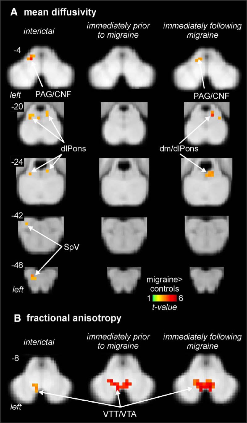
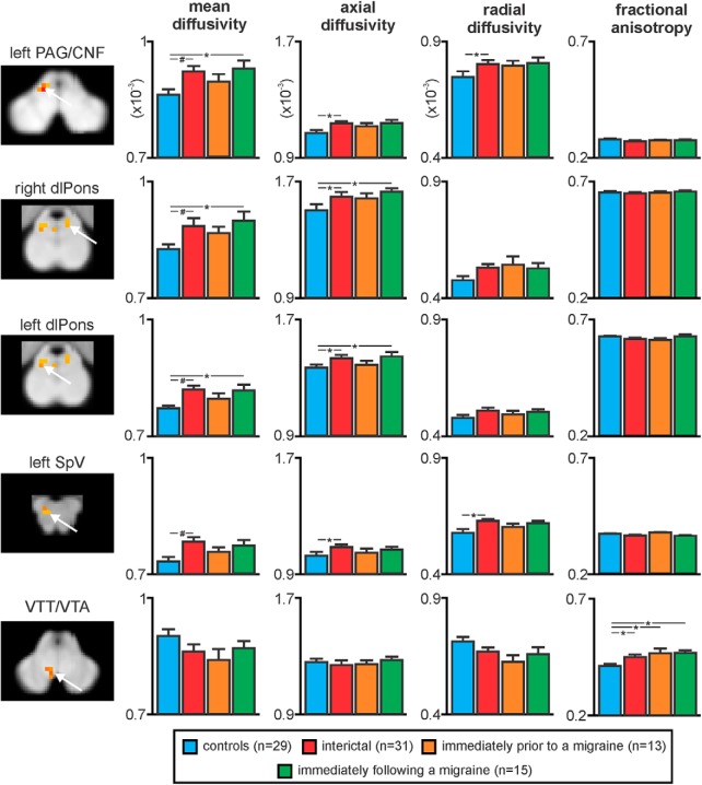
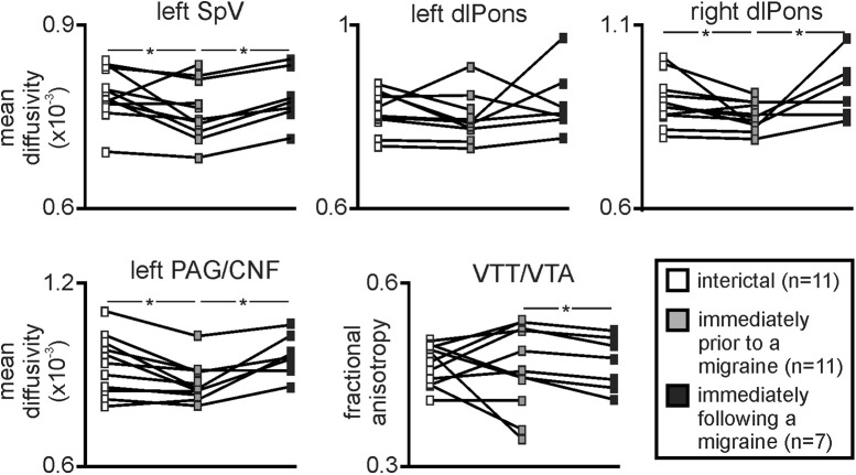
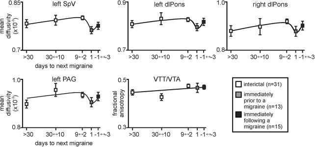
The neural mechanisms responsible for the initiation and expression of migraines remain unknown. Although there is growing evidence of changes in brainstem anatomy and function between attacks, very little is known about brainstem function and structure in the period immediately prior to a migraine. The aim of this investigation is to use brainstem-specific analyses of diffusion weighted images to determine whether the brainstem pain processing regions display altered structure in individuals with migraine across the migraine cycle, and in particular immediately prior to a migraine. Diffusion tensor images (29 controls, 36 migraineurs) were used to assess brainstem anatomy in migraineurs compared with controls. We found that during the interictal phase, migraineurs displayed greater mean diffusivity (MD) in the region of the spinal trigeminal nucleus (SpV), dorsomedial pons (dmPons)/dorsolateral pons (dlPons), and midbrain periaqueductal gray matter (PAG)/cuneiform nucleus (CNF). Remarkably, the MD returned to controls levels during the 24-h period immediately prior to a migraine, only to increase again within the three following days. Additionally, fractional anisotropy (FA) was significantly elevated in the region of the medial lemniscus/ventral trigeminal thalamic tract in migraineurs compared with controls over the entire migraine cycle. These data show that regional brainstem anatomy changes over the migraine cycle, with specific anatomical changes occurring in the 24-h period prior to onset. These changes may contribute to the activation of the ascending trigeminal pathway by either an increase in basal traffic or by sensitizing the trigeminal nuclei to external triggers, with activation ultimately resulting in perception of head pain during a migraine attack.

Keywords: MRI; PAG; brainstem; diffusion tensor imaging; migraine; spinal trigeminal nucleus.


Figures

Figure 1.
Figure 1.
Regional MD (A) and FA differences (B) in migraineurs during the interictal (n = 31), immediately before migraine (n = 13), and immediately following a migraine (n = 15) compared with controls (n = 29). Significantly different clusters are overlaid onto axial brainstem template images and significant increases in migraineurs are represented by a t value with a hot color scale. Slice locations are indicated at the upper left of each axial slice in MNI space. Compared to controls, migraineurs have increased MD during the interictal and immediately following migraine periods in the region of the left SpV, left and right dlPons, and in the region encompassing the PAG/CNF. In addition, migraineurs display an increase in FA in the region of the VTT/VTA during all three phases compared with controls.
Figure 2.
Figure 2.
Plots of mean (±SEM) MD, axial diffusivity, radial diffusivity, and FA values in migraineurs compared with pain-free controls in the left SpV, left and right dlPons, left PAG/CNF, and in the VTT/VTA. Consistent with the voxel-by-voxel analysis, MD was significantly increased during the interictal and immediately following a migraine phase but not immediately before a migraine. In addition, axial diffusivity showed a largely similar pattern of difference in migraineurs whereas radial diffusion was only different during the interictal phase in the PAG/CNF and SpV. In contrast, FA was significantly increased in the area of the VTT/VTA in migraineurs during all three phases; #p < 0.05 voxel-by-voxel analysis; *p < 0.05 two-sample t tests.
Figure 3.
Figure 3.
Plots of MD and FA in 11 migraineurs that were scanned during at least two of the three migraine phases. Note the consistency of MD change which decreases significantly during the period immediately before migraine in the left SpV, right dlPons, and left PAG/CNF. In contrast, FA within the area of the VTT/VTA was significantly greater during the period immediately before compared with immediately following a migraine; *p < 0.05 paired t tests.
Figure 4.
Figure 4.
Plots of mean (±SEM) MD and FA changes in migraineurs over the migraine cycle in the left SpV, left and right dlPons, left PAG/CNF, and in the VTT/VTA. Values are averaged for those migraineurs scanned at least 30 d (n = 18), 30–10 d (n = 6), 9–2 d (n = 7), and 1 d before their next migraine (n = 13), as well as 1–3 d following a migraine (n = 15). Note how MD remains relatively stable over the interictal period and falls dramatically immediately before a migraine before recovering. In contrast, FA remains stable over all three migraine phases.

Similar articles

Cited by

References

    1. Afridi SK, Giffin NJ, Kaube H, Friston KJ, Ward NS, Frackowiak RS, Goadsby PJ (2005) A positron emission tomographic study in spontaneous migraine. Arch Neurol 62:1270–1275. 10.1001/archneur.62.8.1270 - DOI - PubMed
    1. Akerman S, Holland PR, Goadsby PJ (2011) Diencephalic and brainstem mechanisms in migraine. Nat Rev Neurosci 12:570–584. 10.1038/nrn3057 - DOI - PubMed
    1. Alexander AL, Lee JE, Lazar M, Field AS (2007) Diffusion tensor imaging of the brain. Neurotherapeutics 4:316–329. 10.1016/j.nurt.2007.05.011 - DOI - PMC - PubMed
    1. Bahra A, Matharu MS, Buchel C, Frackowiak RS, Goadsby PJ (2001) Brainstem activation specific to migraine headache. Lancet 357:1016–1017. 10.1016/s0140-6736(00)04250-1 - DOI - PubMed
    1. Basser PJ, Pierpaoli C (1996) Microstructural and physiological features of tissues elucidated by quantitative-diffusion-tensor MRI. J Magn Reson B 111:209–219. - PubMed

Publication types