. 2019 Jul 31;6(4):ENEURO.0112-19.2019.
doi: 10.1523/ENEURO.0112-19.2019. Print 2019 Jul/Aug.

Labile Calcium-Permeable AMPA Receptors Constitute New Glutamate Synapses Formed in Hypothalamic Neuroendocrine Cells during Salt Loading

Affiliations

Labile Calcium-Permeable AMPA Receptors Constitute New Glutamate Synapses Formed in Hypothalamic Neuroendocrine Cells during Salt Loading

Shi Di et al. eNeuro. .

Abstract

Magnocellular neuroendocrine cells (MNCs) of the hypothalamus play a critical role in the regulation of fluid and electrolyte homeostasis. They undergo a dramatic structural and functional plasticity under sustained hyperosmotic conditions, including an increase in afferent glutamatergic synaptic innervation. We tested for a postulated increase in glutamate AMPA receptor expression and signaling in magnocellular neurons of the male rat hypothalamic supraoptic nucleus (SON) induced by chronic salt loading. While without effect on GluA1-4 subunit mRNA, salt loading with 2% saline for 5-7 d resulted in a selective increase in AMPA receptor GluA1 protein expression in the SON, with no change in GluA2-4 protein expression, suggesting an increase in the ratio of GluA1 to GluA2 subunits. Salt loading induced a corresponding increase in EPSCs in both oxytocin (OT) and vasopressin (VP) neurons, with properties characteristic of calcium-permeable AMPA receptor-mediated currents. Unexpectedly, the emergent AMPA synaptic currents were silenced by blocking protein synthesis and mammalian target of rapamycin (mTOR) activity in the slices, suggesting that the new glutamate synapses induced by salt loading require continuous dendritic protein synthesis for maintenance. These findings indicate that chronic salt loading leads to the induction of highly labile glutamate synapses in OT and VP neurons that are comprised of calcium-permeable homomeric GluA1 AMPA receptors. The glutamate-induced calcium influx via calcium-permeable AMPA receptors would be expected to play a key role in the induction and/or maintenance of activity-dependent synaptic plasticity that occurs in the magnocellular neurons during chronic osmotic stimulation.

Keywords: excitability; osmoregulation; oxytocin; paraventricular; supraoptic; vasopressin.


Figures

None
Graphical abstract
Figure 1.
Figure 1.
Effect of salt loading on VP mRNA expression in SON. A, Image of a Nissl-stained coronal section of rat brain at the level of the hypothalamus showing the approximate locations of the 1-mm SON punches (dashed circles). B, The relative VP mRNA expression measured by qPCR in SON punches from control (control) and salt-loaded (NaCl) rats, normalized to control; *p < 0.05.
Figure 2.
Figure 2.
Effects of chronic salt loading on AMPA receptor subunit expression in the SON. A, qPCR of AMPA receptor subunit mRNA expression in SON punches from control and salt-loaded (NaCl) rats revealed no significant change in any of the AMPA receptor subunit mRNAs, GluA1-4, with salt loading. B1, Representative Western blots of GluA1 and GluA2 total protein expression in SON punches from four control rats (C1–C4) and five (GluA1: Na1–Na5) or four (GluA2: Na1–Na4) salt-loaded rats. B2, Mean optical density measurements of GluA1 and GluA2 protein bands from SON tissue from control (N = 8) and salt-loaded (NaCl, N = 7) rats revealed an increase in total GluA1 protein with salt loading. C, Surface and intracellular GluA1 and GluA2 protein levels in SON punches from control (N = 7, 8, respectively) and salt-loaded (NaCl, N = 5, 7, respectively) rats determined by Western blot following BS3 surface protein crosslinking. Inset, Representative Western blots of SON tissue from a control rat treated with the membrane impermeant crosslinking agent BS3 and probed with antisera to GluA2 and β-actin. Two GluA2 bands were detected: a high molecular weight band representing the crosslinked surface protein aggregate (S) and a band at the predicted molecular weight for GluA2 (∼100 kDa) that represents the unmodified intracellular pool (I). β-Actin served as an intracellular control and was found at its predicted molecular weight (∼40 kDa), confirming the lack of crosslinking of intracellular proteins. D, Surface-to-intracellular (S/I) protein ratios determined following BS3 surface protein crosslinking for GluA1-GluA4 AMPA receptor subunits in SON punches from control and salt-loaded (NaCl) rats. Despite the increase in total GluA1 protein, there was no change in the subunit S/I ratios between SON tissues from control and salt-loaded rats for any of the AMPA receptor subunits; *p < 0.05.
Figure 3.
Figure 3.
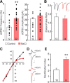
Increase in inwardly rectifying AMPA receptor signaling following salt loading. A, Scatter plots of individual neuronal means and group means of sEPSC frequencies and amplitudes. Identified VP neurons recorded in slices from salt-loaded VP-eGFP rats (NaCl) showed a significant increase in the mean sEPSC frequency and amplitude compared to the control group. B, The paired-pulse ratio of eEPSCs (inset) was unchanged following salt loading, suggesting that the increased glutamatergic synaptic activity was not due to a change in the probability of glutamate release. C, The eEPSC current−voltage relationship showed inward rectification at Vm > 0 mV in SON neurons from salt-loaded rats (red curve, n = 6) but not controls (black curve, n = 4). The mean I-V curves are group averages of normalized individual I-V curves, expressed as percent of eEPSC recorded at −60 mV. D, Representative AMPA receptor-mediated eEPSC responses recorded at holding potentials of –60 mV and +40 mV in SON neurons from control and salt-loaded rats. E, The rectification index (eEPSC amplitude at –60 mV/eEPSC amplitude at +40 mV) increased in SON neurons from salt-loaded rats, suggesting the emergence of calcium-permeable AMPA receptor-containing glutamate synapses with salt loading. VP and OT neurons were pooled together for panels B–E. Numerals in bars represent recorded cell numbers; *p < 0.05, **p < 0.01. Scale bars = 100 pA, 20 ms.
Figure 4.
Figure 4.
Change in EPSC sensitivity to the calcium-permeable AMPA receptor antagonist NAS following salt loading. A, NAS reduced the mean frequency (left) and amplitude (right) of sEPSCs over time in SON neurons from salt-loaded (NaCl) rats, but not in SON neurons from control rats. B, The effect of NAS on mean sEPSC frequency (Freq), amplitude (Amp), and decay time (Decay) in SON neurons from salt-loaded rats (NaCl) and control rats, expressed as a percentage of baseline. NAS decreased the sEPSC frequency and amplitude in SON neurons from salt-loaded rats but not control rats. C, NAS caused a decrease in the mean amplitude of eEPSCs in SON neurons from salt-loaded rats, but not control rats. NAS had no effect on the paired-pulse ratio of eEPSCs (PPR) in SON neurons from either control or salt-loaded rats. D, NAS had no effect on the eEPSC rectification index in SON neurons from control rats, but reversed the increase in the rectification index in SON neurons from salt-loaded rats. Numerals in bars represent recorded cell numbers; *p < 0.05, **p < 0.01 compared to control or baseline.
Figure 5.
Figure 5.
Correlation of the membrane surface area to NAS sensitivity. There was a significant correlation between the increase in magnocellular neuron surface area, as measured by membrane capacitance (Cm), and the change in eEPSC amplitude in NAS.
Figure 6.
Figure 6.
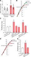
The maintenance of new inwardly rectifying AMPA currents is dependent on protein synthesis. A, Blocking protein synthesis with CHX (25 μM) had no effect on sEPSC frequency in SON neurons from control rats, but reversed the increase in sEPSC frequency induced in SON neurons by salt loading to the control baseline level (one-way ANOVA followed by Dunnett’s test). B, Blocking protein synthesis with CHX (NaCl+ CHX) eliminated the increase in eEPSC inward rectification seen in SON neurons from salt-loaded rats (NaCl) compared to SON neurons from control rats. C, CHX had no effect on the rectification index in eEPSCs in SON neurons from control rats (control+CHX), but reversed the increase in the rectification index (NaCl+CHX) and occluded the effect of NAS (NaCl+CHX+NAS) in SON neurons from salt-loaded rats. Inhibition of gene transcription with ACT (25 μM) had no effect on the eEPSC rectification index (NaCl+ACT) and did not prevent the NAS-induced reduction in the rectification index (NaCl+ACT+NAS) in neurons from salt-loaded rats. D, E, Blocking mTOR activity with rapamycin (NaCl+Rapa) blocked the eEPSC inward rectification induced by salt loading and returned the eEPSC rectification index to the control level (one-way ANOVA followed by Dunnett’s test); *p < 0.05.

Similar articles

Cited by

References

    1. Al-Ghoul WM, Meeker RB, Greenwood RS (1997) Differential expression of five N-methyl-D-aspartate receptor subunit mRNAs in vasopressin and oxytocin neuroendocrine cells. Brain Res Mol Brain Res 44:262–272. - PubMed
    1. Arnauld E, Wattiaux JP, Arsaut J, Rostène W, Vincent JD (1993) Alterations in vasopressin and oxytocin messenger RNA in the rat supraoptic nucleus during dehydration-rehydration evaluated by in situ hybridization and northern blotting. Neurosci Lett 149:177–181. 10.1016/0304-3940(93)90765-d - DOI - PubMed
    1. Bonfanti L, Theodosis DT (2009) Polysialic acid and activity-dependent synapse remodeling. Cell Adh Migr 3:43–50. 10.4161/cam.3.1.7258 - DOI - PMC - PubMed
    1. Borges K, Dingledine R (1998) AMPA receptors: molecular and functional diversity. Prog Brain Res 116:153–170. - PubMed
    1. Boudaba C, Di S, Tasker JG (2003) Presynaptic noradrenergic regulation of glutamate inputs to hypothalamic magnocellular neurones. J Neuroendocr 15:803–810. 10.1046/j.1365-2826.2003.01063.x - DOI - PubMed

Publication types

LinkOut - more resources