. 2019 Jul 12;10(1):3094.
doi: 10.1038/s41467-019-10910-w.

AMPA receptor GluA2 subunit defects are a cause of neurodevelopmental disorders

Vincenzo Salpietro  1   2   3 Christine L Dixon  4 Hui Guo  5   6 Oscar D Bello  4 Jana Vandrovcova  1 Stephanie Efthymiou  1   4 Reza Maroofian  1 Gali Heimer  7 Lydie Burglen  8 Stephanie Valence  9 Erin Torti  10 Moritz Hacke  11 Julia Rankin  12 Huma Tariq  1 Estelle Colin  13   14 Vincent Procaccio  13   14 Pasquale Striano  2   3 Kshitij Mankad  15 Andreas Lieb  4 Sharon Chen  16 Laura Pisani  16 Conceicao Bettencourt  17 Roope Männikkö  1 Andreea Manole  1 Alfredo Brusco  18 Enrico Grosso  18 Giovanni Battista Ferrero  19 Judith Armstrong-Moron  20 Sophie Gueden  21 Omer Bar-Yosef  7 Michal Tzadok  7 Kristin G Monaghan  10 Teresa Santiago-Sim  10 Richard E Person  10 Megan T Cho  10 Rebecca Willaert  10 Yongjin Yoo  22 Jong-Hee Chae  23 Yingting Quan  6 Huidan Wu  6 Tianyun Wang  5   6 Raphael A Bernier  24 Kun Xia  6 Alyssa Blesson  25 Mahim Jain  25 Mohammad M Motazacker  26 Bregje Jaeger  27 Amy L Schneider  28 Katja Boysen  28 Alison M Muir  29 Candace T Myers  30 Ralitza H Gavrilova  31 Lauren Gunderson  31 Laura Schultz-Rogers  31 Eric W Klee  31 David Dyment  32 Matthew Osmond  32   33   34 Mara Parellada  35 Cloe Llorente  36 Javier Gonzalez-Peñas  37 Angel Carracedo  38   39 Arie Van Haeringen  40 Claudia Ruivenkamp  40 Caroline Nava  41 Delphine Heron  41 Rosaria Nardello  42 Michele Iacomino  43 Carlo Minetti  2   3 Aldo Skabar  44 Antonella Fabretto  44 SYNAPS Study GroupMiquel Raspall-Chaure  45 Michael Chez  46 Anne Tsai  47 Emily Fassi  48 Marwan Shinawi  48 John N Constantino  49 Rita De Zorzi  50 Sara Fortuna  50 Fernando Kok  51   52 Boris Keren  41 Dominique Bonneau  13   14 Murim Choi  22 Bruria Benzeev  7 Federico Zara  43 Heather C Mefford  29 Ingrid E Scheffer  28 Jill Clayton-Smith  53   54 Alfons Macaya  45 James E Rothman  4   55 Evan E Eichler  5   56 Dimitri M Kullmann  57 Henry Houlden  58
Collaborators, Affiliations

AMPA receptor GluA2 subunit defects are a cause of neurodevelopmental disorders

Vincenzo Salpietro et al. Nat Commun. .

Abstract

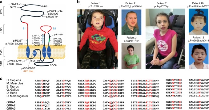
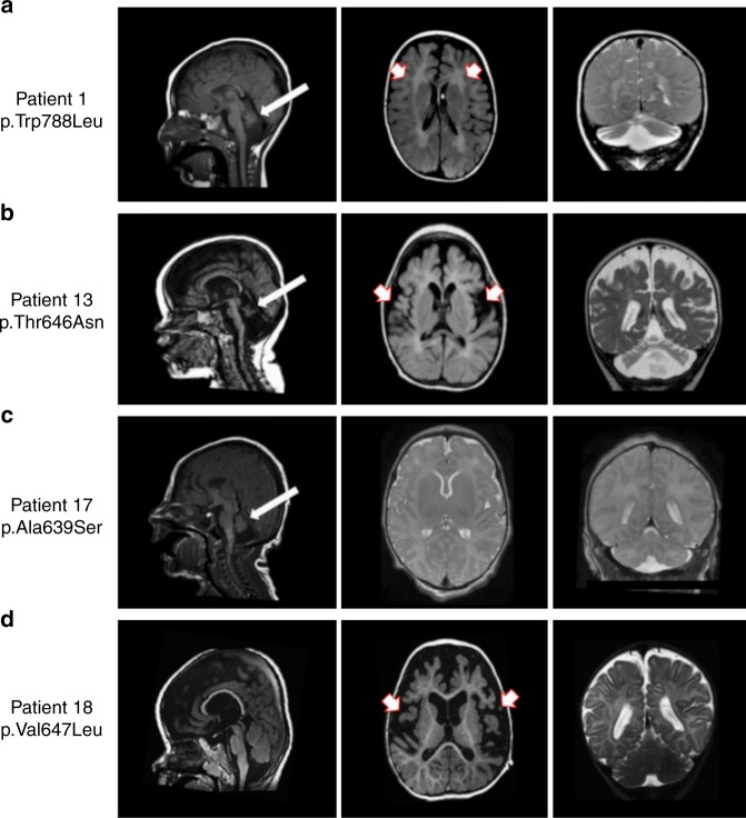
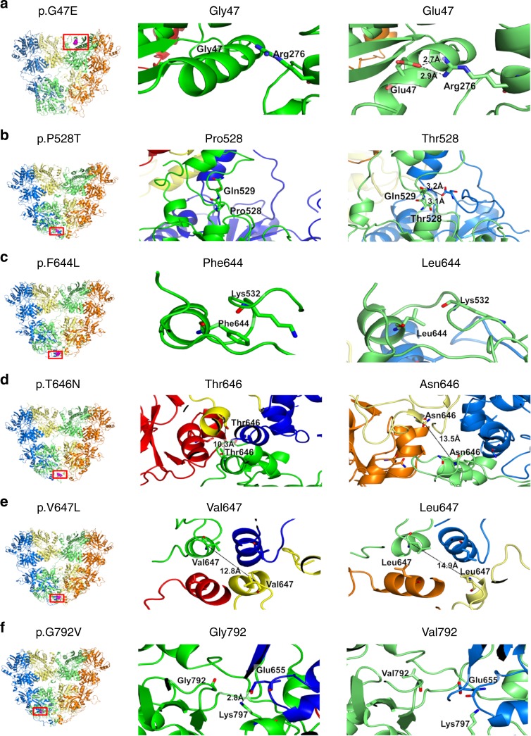
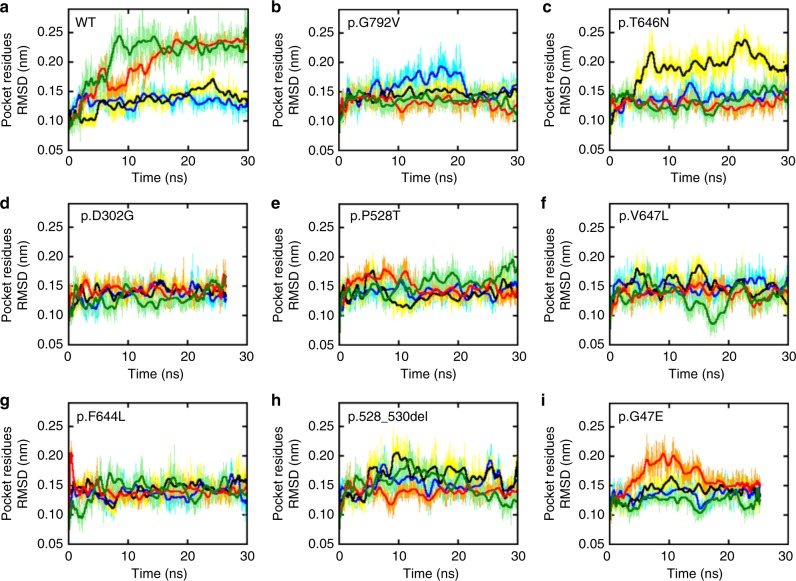
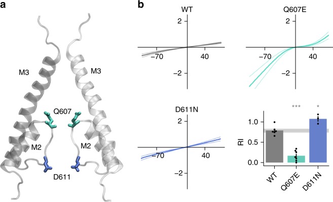
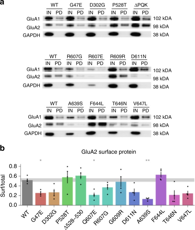
AMPA receptors (AMPARs) are tetrameric ligand-gated channels made up of combinations of GluA1-4 subunits encoded by GRIA1-4 genes. GluA2 has an especially important role because, following post-transcriptional editing at the Q607 site, it renders heteromultimeric AMPARs Ca2+-impermeable, with a linear relationship between current and trans-membrane voltage. Here, we report heterozygous de novo GRIA2 mutations in 28 unrelated patients with intellectual disability (ID) and neurodevelopmental abnormalities including autism spectrum disorder (ASD), Rett syndrome-like features, and seizures or developmental epileptic encephalopathy (DEE). In functional expression studies, mutations lead to a decrease in agonist-evoked current mediated by mutant subunits compared to wild-type channels. When GluA2 subunits are co-expressed with GluA1, most GRIA2 mutations cause a decreased current amplitude and some also affect voltage rectification. Our results show that de-novo variants in GRIA2 can cause neurodevelopmental disorders, complementing evidence that other genetic causes of ID, ASD and DEE also disrupt glutamatergic synaptic transmission.


Conflict of interest statement

E.T., K.G.M., T.S.-S., R.E.P., R.W., and M.T.C. are employees of GeneDx. E.E.E. is on the scientific advisory board (SAB) of DNAnexus, Inc. The remaining authors declare no competing interests.

Figures

Fig. 1
Fig. 1
GRIA2 intragenic de-novo variants identified in this study. a Schematic of the human GluA2 protein (NP_000817.2) indicating the positions of twelve missense changes (dot arrows), two frameshift deletions (cross arrows), two splice-site variants (arrows) and an in-frame deletion (dot arrow). Glutamate binding regions are displayed in red, flip/flop alternatively spliced region is represented in green. b Left panel: Patient 1, carrying the de-novo p.W788L GluA2Flop variant, at 3 years, exhibitinghypotonia and an oculogyric crisis; he is wheelchair dependent. Middle left panel: Patient 2 (top) carrying the de-novo p.P528_K530del in-frame deletion, at 12 years; Patient 3 (bottom) carrying the de-novo p.D611N variant, at 18 years, exhibiting hand-wringing suggestive of RTT. Middle right panel: Patient 7, carrying the de-novo p.Q607E/p.R607G heterozygous mutation (affecting the Q/R editing site) at 10 years, exhibiting hand-wringing as part of a RTT-like presentation. Right panel: Patient 10 (top) carrying the de-novo p.F595LfsX37 variant, at 5 years; Patient 12 (bottom) carrying the de-novo p.P286LfsX14 at 6 years. c Multiple alignment showing GluA2 protein complete conservation across species and inter AMPAR homolog subunits (GluA1, GluA3, and GluA4) alignment. Human GRIA2 (NP_000817.2), mouse GRIA2 (NP_001077275.1), bos taurus GRIA2 (NP_001069789.2), gallus gallus GRIA2 (NP_001001775.2), danio rerio (NP_571970.2), drosophila melanogaster (NP_476855.1), Human GRIA1 (NP_000818.2) Human GRIA3 (NP_015564.4), and Human GRIA4 (NP_000820.3)
Fig. 2
Fig. 2
Brain imaging in 4 individuals with GRIA2-related DEE and brain and cerebellar atrophy. a Sagittal T1 weighted (left panel), Axial T1 weighted (middle panel), and coronal T2 weighted (right panel) images from Patient 1 (carrying the de-novo p.W788L mutation) scanned at the age of 3 years. There is reduction in the cerebral white matter (red and white arrows), brain volume and marked cerebellar atrophy with vermian deficiency (white arrows). b Sagittal T1 weighted (left panel) images from Patient 13 (carrying the de-novo p.T646N mutation) at the age of 2 months, and axial T1 weighted (middle panel) and coronal T2 weighted images (right panel) from the same Patient at the age of 11 months demonstrate white matter signal abnormality (white and red arrows) with generalized reduction in the cerebral white matter volume and cerebellar atrophy with vermian deficiency (white arrows). c Sagittal T1 weighted (left panel), Axial T2 weighted (middle panel) and coronal T2 weighted (right panel) images from Patient 17 (carrying the de-novo p. A639S mutation) at the age of 6 days; there is an underdeveloped/hypoplastic cerebellum (white arrows) and delayed maturation of myelin. d Sagittal T1 weighted (left panel), axial T1 weighted (middle panel) and coronal T2 weighted (right poanel) images from Patient 18 (carrying the de-novo p.V647L mutation) scanned at the age of 18 months; there is a global cerebral atrophy and white matter changes which suggest hypomyelination (white and red arrows). Although the volume of the cerebellar hemispheres is preserved, atrophy of the inferior cerebellar vermis and wide cerebellar sulci are seen
Fig. 3
Fig. 3
Molecular modeling and predicted consequences of 6 GRIA2 de-novo missense variants Left panel: Six modeled GRIA2 de-novo missense variants (highlighted in purple within red rectangle) affecting the amino-terminal domain (ATD) and ligand-binding domain (LBD) of GluA2; Middle panel: particular of the wild-type GlUA2 residue; Right panel: particular of the mutated GluA2 residue. a For variant p.G47E, the presence of the glutamic acid residue (right panel) in place of the glycine residue (middle panel) is predicted to cause the formation of two hydrogen bonds with a neighboring arginine residue; hydrogen bond distances are shown for the mutant structure. b The presence of a Threonine residue in position 528 (right panel) is predicted to cause a slight change in the backbone conformation of the neighboring residues that, in turn, allows hydrogen bonding formation between the two chains forming the LBD domain. c Mutation p.F644L causes the loss of hydrophobic interactions between residue 644 and the side chain of Lys532. d For mutation p.T646N, the presence of a more hydrophilic Asparagine residue increases the distance between the distal chains by about 3 Å, at the interface of LBD and TMD (right panel). e For mutation p.V647L, increased hydrophobicity of Leucine (right panel) compared to Valine (middle panel) increases the separation of helices at the interface between LBD and TMD by about 2 Å. f For variant p.G792V, the presence of a more hydrophobic Valine residue in close proximity to the binding site is predicted to cause a sliding movement with respect to the neighboring chain, disrupting the interchain salt bridge between Glu655 and Lys797; hydrogen bond distance is shown in the wild-type GluA2 (middle panel)
Fig. 4
Fig. 4
GluA2 Molecular dynamic stimulations predict reduced mobility at the agonist binding site. Root Mean Square Deviation (RMSD) of the GluA2 Glutamate binding pocket (GLU) amino acids along the simulated time for the wild-type protein (top left) and GRIA2 mutants (ai). In all panels the color code is: chain A (blue/cyan), chain B (black/yellow), chain C (red/orange), and chain D (light/dark green). Thicker lines indicate running averages over 100 samples
Fig. 5
Fig. 5
Agonist-evoked currents in HEK cells. 1 mM KA was applied transiently to HEK cells expressing GluA2 and stargazin (a), or GluA1, GluA2 and stargazin (b). Amplitude was compared to WT (*p < 0.05, **p < 0.01, ***p < 0.001) and for co-expression also to the negative control (GluA1 without GluA2, —, #p < 0.05, ##p < 0.01, ###p < 0.001). Mean ± SEM. Data are from the following numbers of independent cells: (a) WT:25, G47E:7, D302G:6, P528T:7, Δ528–530:11, Q607E:10, G609R:13, D611N:8, A639S:10, F644L:6, T646N:10, V647L:10 (b) —:21, WT:28, G47E:7, D302G:10, P528T:9, Δ528–530:15, Q607E:12, G609R:11, D611N:15, A639S:19, F644L:14, T646N:12, and V647L:6
Fig. 6
Fig. 6
Rectification of GluA2 mutants when co-expressed with GluA1. Voltage was ramped from −104 mV to +76 in order to assess rectification of KA-evoked currents. Ramp currents recorded in the absence of KA were subtracted from ramp currents in 1 mM KA. Controls showed linear current–voltage (I–V) relations for cells co-expressing GluA1 and GluA2 WT, and clear rectification in cells transfected with GluA2 alone. The panels show average ramps ±SEM for each mutant (n ≥ 6 cells per mutant). Rectification index (RI) was quantified as (I+40/I−70) *(−7/4). RI was compared to WT (*p < 0.05, **p < 0.01, ***p < 0.001) and GluA1-negative control (#p < 0.05, ##p < 0.01, ###p < 0.001). Data are averaged from the following numbers of cells per mutant: —:19, WT:19, G47E:6, D302G:7, P528T:9, Δ528–530:14, Q607E:10, G609R:11, D611N:13, A639S:18, F644L:10, T646N:10, V647L:5
Fig. 7
Fig. 7
Changes at the polyamine binding site alter rectification in GluA2 homomeric channels. a Pore detail of 2 opposing subunits in GluA2 tetramer (cryo-EM structure pdb 6dm0). b Ramps were applied to HEK cells expressing homomeric GluA2 channels and rectification quantified as for Fig. 4 (*p < 0.05, **p < 0.01, ***p < 0.001). Number of cells recorded per mutant: WT:6, Q607E:7, D611N:3
Fig. 8
Fig. 8
Scatter plot summarizing changes in current amplitude and rectification when GluA2 is co-expressed with GluA1 in HEK cells. Relationship between RI and KA-evoked current amplitude for mutant GluA2, compared to WT and negative (GluA1) control (—). Error bars are SEM
Fig. 9
Fig. 9
Some GluA2 mutations disrupt surface expression. a Samples of transfected HEK cells biotinylated before (IN) and after purification by pull down with streptavidin beads (PD). b IN and PD were quantified relative to GADPH and then normalized to a WT control on the same gel. Surface protein was quantified as PD/IN. *p < 0.05, **p < 0.01 vs WT. N = 4 experiments from independent batches of transfected cells

Comment in

References

    1. Heyne HO, et al. De-novo variants in neurodevelopmental disorders with epilepsy. Nat. Genet. 2018;50:1048–1053. doi: 10.1038/s41588-018-0143-7. - DOI - PubMed
    1. Chen S, et al. An interactome perturbation framework prioritizes damaging missense mutations for developmental disorders. Nat. Genet. 2018;50:1032–1040. doi: 10.1038/s41588-018-0130-z. - DOI - PMC - PubMed
    1. Deciphering Developmental Disorders Study. Prevalence and architecture of de-novo mutations in developmental disorders. Nature. 2017;542:433–438. doi: 10.1038/nature21062. - DOI - PMC - PubMed
    1. Lombardi LM, Baker SA, Zoghbi HY. MECP2 disorders: from the clinic to mice and back. J. Clin. Invest. 2015;125:2914–2923. doi: 10.1172/JCI78167. - DOI - PMC - PubMed
    1. Scheffer IE, et al. ILAE classification of the epilepsies: position paper of the ILAE Commission for Classification and Terminology. Epilepsia. 2017;58:512–521. doi: 10.1111/epi.13709. - DOI - PMC - PubMed

Publication types

Substances