. 2019 Jul 12;9(1):10121.
doi: 10.1038/s41598-019-46568-z.

In vivo two-photon microscopy of the human eye

Affiliations

In vivo two-photon microscopy of the human eye

Francisco J Ávila et al. Sci Rep. .

Abstract

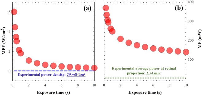
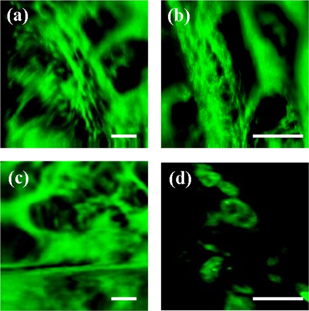
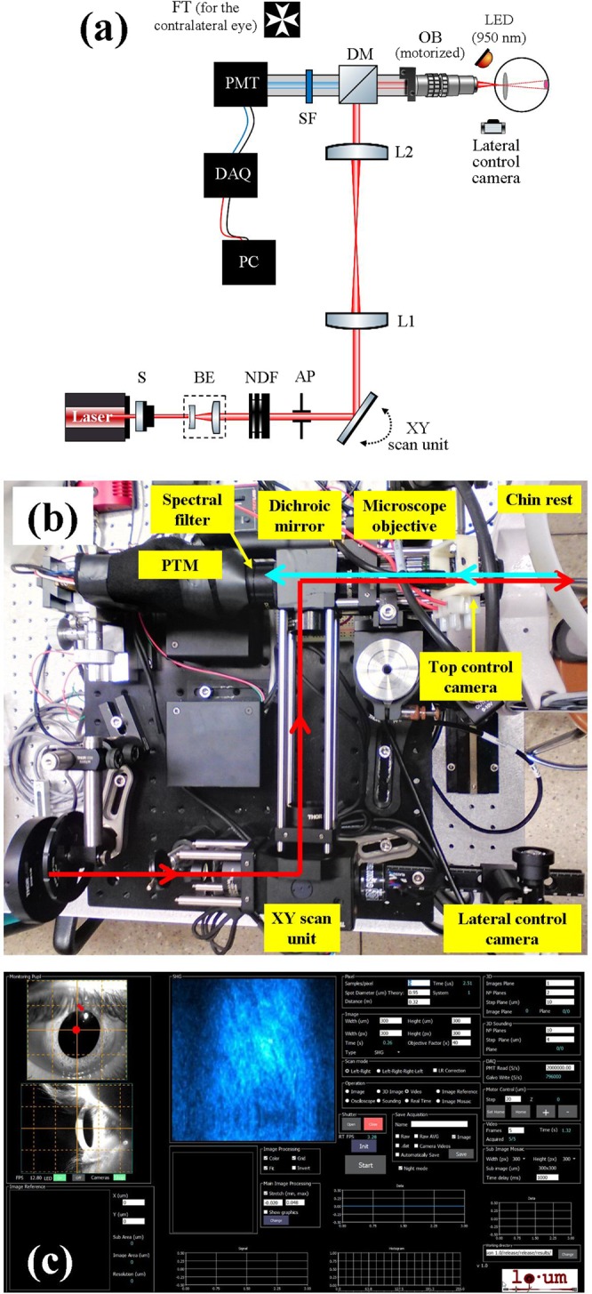
Two-photon (2P) microscopy is a powerful tool for imaging and exploring label-free biological tissues at high resolution. Although this type of microscopy has been demonstrated in ex vivo ocular tissues of both humans and animal models, imaging the human eye in vivo has always been challenging. This work presents a novel compact 2P microscope for non-contact imaging of the anterior part of the living human eye. The performance of the instrument was tested and the maximum permissible exposure to protect ocular tissues established. To the best of our knowledge, 2P images of the in vivo human cornea, the sclera and the trabecular meshwork are shown for the very first time. Acquired images are of enough quality to visualize collagen arrangement and morphological features of clinical interest. Future implementations of this technique may constitute a potential tool for early diagnosis of ocular diseases at submicron scale.


Conflict of interest statement

The authors declare no competing interests.

Figures

Figure 1
Figure 1
(a) MPE as a function of exposure time to protect the cornea during 2P imaging. (b) Maximum laser power to protect the human retina as a function of the exposure time (this was computed for a fixed retinal area of 7.6 mm2; see details in the text).
Figure 2
Figure 2
(ad) SHG images of the in vivo corneal stroma (apex region) of two different subjects (#1 (a, c); #2 (b,d) and two different depth locations: (a,b) anterior stroma ~50-µm depth; (c,d) posterior stroma ~350-µm depth. (e,f) Comparison of SHG images of the corneal stroma recorded in in vivo and ex vivo conditions, where the spatial pattern could be clearly visualized. The arrows indicate the two POs. Scale bar: 50 µm. The image corresponding to the ex vivo sample was recorded as explained in ref.. (g,h) PO histograms showing the two orientations of the fibers (peaks). Each in vivo SHG image corresponds to an individual frame (image averaging was not used). Scale bar: 50 µm.
Figure 3
Figure 3
(ad) SHG images of the living human sclera at two different depth locations spaced ~50-µm in subjects #1 (a,c) and #2 (b,d). (eh) SHG images of the living human cornea (e,f) and sclera (g,h) recorded 2 s apart. Bar length: 50 µm.
Figure 4
Figure 4
2P images (combining TPEF and SHG signals) of the trabecular meshwork region for subjects #1 (a,b) and #2 (c). (d) Shows details of the juxta-canalicular tissue. Scale bar: 50 µm.
Figure 5
Figure 5
(a) Schematic diagram of the compact 2P microscope. S, shutter; BE, beam expander; NDF, neutral density filter; AP, aperture; L1 and L2, achromatic doublets; DM, dichroic mirror; OB, microscope objective; SF, spectral filter; PMT, photomultiplier tube; DAQ, data acquisition card; PC, computer. (b) Picture showing a top view of the actual instrument. The main elements are highlighted. (c) Customized user’s interface used for eye centration and image recording. The operator can choose the dimensions of the scanning area, the image size (in pixels) and the exposure time. For optical sectioning, the user must also set the number of planes and the axial distance between correlative planes.
Figure 6
Figure 6
Simplified schematic indicating the locations of the imaged areas. It should be notice that this figure is intended for illustration, as the objective is unique and the eye rotates within its own orbit for the incident beam to be always perpendicular to the imaged region. For simplicity the eye’s rotation was omitted here.

References

    1. Maurice DM. The structure and transparency of the cornea. J Physiol. 1957;136:263–286. doi: 10.1113/jphysiol.1957.sp005758. - DOI - PMC - PubMed
    1. Guthoff RF, Zhivov A, Stachs O. In vivo confocal microscopy, an inner vision of the cornea - A major review. Clin. Exp. Ophthalmol. 2009;37:100–117. doi: 10.1111/j.1442-9071.2009.02016.x. - DOI - PubMed
    1. Han, S. B., Liu, Y. C., Noriega, K. M. & Mehta, J. S. Applications of anterior segment optical coherence tomography in cornea and ocular surface diseases. J. Ophthalmol. 4971572 (2016). - PMC - PubMed
    1. Denk W, Strickler JH, Webb WW. Two-photon laser scanning fluorescence microscopy. Science. 1990;248:73–76. doi: 10.1126/science.2321027. - DOI - PubMed
    1. Ustine A, Piston DW. A simple introduction to multiphoton microscopy. J. Microsc. 2011;243:221–226. doi: 10.1111/j.1365-2818.2011.03532.x. - DOI - PubMed

Publication types

MeSH terms