. 2019 Jul 12;9(1):10142.
doi: 10.1038/s41598-019-46505-0.

Differential effects of slow rewarming after cerebral hypothermia on white matter recovery after global cerebral ischemia in near-term fetal sheep

Affiliations

Differential effects of slow rewarming after cerebral hypothermia on white matter recovery after global cerebral ischemia in near-term fetal sheep

V Draghi et al. Sci Rep. .

Abstract

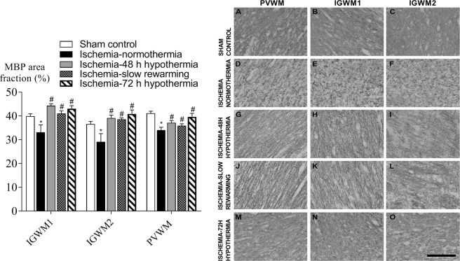
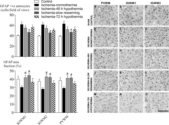
It is widely believed that rewarming slowly after therapeutic hypothermia for hypoxic-ischemic (HI) encephalopathy can improve outcomes, but its impact on white matter injury after HI is unclear. Fetal sheep (0.85 gestation) received 30 min ischemia-normothermia (n = 8), or hypothermia from 3-48 h with rapid spontaneous rewarming over 1 h (ischemia-48 h hypothermia, n = 8), or 48 h with slow rewarming over 24 h (ischemia-slow rewarming, n = 7) or 72 h with rapid rewarming (ischemia-72 h hypothermia, n = 8). Ischemia was associated with loss of total and mature oligodendrocytes and reduced area fraction of myelin basic protein (MBP) and 2',3'-cyclic nucleotide 3'-phosphodiesterase (CNPase; immature/mature oligodendrocytes) and increased microglia and astrocytes. Total numbers of oligodendrocytes were increased by all hypothermia protocols but only ischemia-72 h hypothermia attenuated loss of mature oligodendrocytes. All hypothermia protocols similarly increased the area fraction of MBP, whereas there was only an intermediate effect on the area fraction of CNPase. Microglia were suppressed by all hypothermia protocols, with the greatest reduction after ischemia-72 h hypothermia, and an intermediate effect after ischemia-slow rewarming. By contrast, induction of astrocytes was significantly reduced only after ischemia-slow rewarming. In conclusion, slow rewarming after hypothermia did not improve oligodendrocyte survival or myelination or suppression of microgliosis compared to fast rewarming, but modestly reduced astrocytosis.


Conflict of interest statement

The authors declare no competing interests.

Figures

Figure 1
Figure 1
Total, Olig-2-positive, oligodendrocyte number (left) and photomicrographs (right) in the intragyral white matter of the first (B,E,H,K,N) and second (C,F,I,L,O) parasagittal gyri and periventricular (A,D,G,J,M) white matter in the sham control (A–C, n = 9), ischemia-normothermia (D–F, n = 8), ischemia-48 h hypothermia (G–I, n = 8), ischemia-slow rewarming (JL, n = 7) and ischemia-72 h hypothermia (M–O, n = 8) groups. Data are mean ± SEM. *P < 0.05 vs sham control. #P < 0.05 vs ischemia-normothermia. Scale bar 200 µm.
Figure 2
Figure 2
Mature, CC-1-positive, oligodendrocyte number (left) and photomicrographs (right) in the intragyral white matter of the first (B,E,H,K,N) and second (C,F,I,L,O) parasagittal gyri and periventricular (A,D,G,J,M) white matter in the sham control (AC, n = 9), ischemia-normothermia (DF, n = 8), ischemia-48 h hypothermia (G–I, n = 8), ischemia-slow rewarming (JL, n = 7) and ischemia-72 h hypothermia (MO, n = 8) groups. Data are mean ± SEM. *P < 0.05 vs sham control. #P < 0.05 vs ischemia-normothermia. Scale bar 200 µm.
Figure 3
Figure 3
Area fraction of myelin basic protein (MBP, left) and photomicrographs (right) in the intragyral white matter of the first (B,E,H,K,N) and second (C,F,I,L,O) parasagittal gyri and periventricular (A,D,G,J,M) white matter in the sham control (A–C, n = 9), ischemia-normothermia (D–F, n = 8), ischemia-48 h hypothermia (G–I, n = 8), ischemia-slow rewarming (J-L, n = 7) and ischemia-72 h hypothermia (M–O, n = 8) groups. Data are mean ± SEM. *P < 0.05 vs sham control. #P < 0.05 vs ischemia-normothermia. Scale bar 200 µm.
Figure 4
Figure 4
Area fraction of 2′,3′-cyclic nucleotide 3′-phosphodiesterase (CNPase, left) and photomicrographs (right) in the intragyral white matter of the first (B,E,H,K,N) and second (C,F,I,L,O) parasagittal gyri and periventricular (A,D,G,J,M) white matter in the sham control (A–C, n = 9), ischemia-normothermia (D–F, n = 8), ischemia-48 h hypothermia (G–I, n = 8), ischemia-slow rewarming (J–L, n = 7) and ischemia-72 h hypothermia (M–O, n = 8) groups. Data are mean ± SEM. *P < 0.05 vs sham control. Scale bar 200 µm.
Figure 5
Figure 5
Ionized calcium-binding adapter molecule 1 (Iba-1) positive microglial number (left) and photomicrograph (right) in the periventricular white matter (A,D,G,J,M) and the intragyral white matter of the first  (B,E,H,K,N) and second parasagittal gyri (C,F,I,L,O) in the sham control (AC, n = 9), ischemia-normothermia (DF, n = 8), ischemia-48 h hypothermia (GI, n = 8), ischemia-slow rewarming (J-L, n = 7) and ischemia-72 h hypothermia (MO, n = 8) groups. Data are mean ± SEM. *P < 0.05 vs sham control. #P < 0.05 vs ischemia-normothermia. a P < 0.05 vs ischemia-48 h hypothermia. Scale bar 200 µm.
Figure 6
Figure 6
Glial-fibrillary-acidic protein (GFAP)-positive astrocyte number (top left) and area fraction (bottom left) and photomicrographs (right) in the periventricular white matter (A,D,G,J,M) and the intragyral white matter of the first (B,E,H,K,N) and second parasagittal gyri (C,F,I,L,O) in the sham control (AC, n = 9), ischemia-normothermia (DF, n = 8), ischemia-48 h hypothermia (GI, n = 8), ischemia-slow rewarming (JL, n = 7) and ischemia-72 h hypothermia (MO, n = 8) groups. Data are mean ± SEM. *P < 0.05 vs sham control. #P < 0.05 vs ischemia-normothermia. Scale bar 200 µm.

References

    1. Edwards AD, et al. BMJ (Clinical research ed.) 2010. Neurological outcomes at 18 months of age after moderate hypothermia for perinatal hypoxic ischaemic encephalopathy: synthesis and meta-analysis of trial data; p. c363. - PMC - PubMed
    1. Shankaran S, et al. Neonatal Magnetic Resonance Imaging Pattern of Brain Injury as a Biomarker of Childhood Outcomes following a Trial of Hypothermia for Neonatal Hypoxic-Ischemic Encephalopathy. J. Pediatr. 2015;167:987–993 e983. doi: 10.1016/j.jpeds.2015.08.013. - DOI - PMC - PubMed
    1. Wassink G, et al. A working model for hypothermic neuroprotection. J. Physiol. 2018;596:5641–5654. doi: 10.1113/JP274928. - DOI - PMC - PubMed
    1. Davidson JO, et al. How long is sufficient for optimal neuroprotection with cerebral cooling after ischemia in fetal sheep? J. Cereb. Blood Flow Metab. 2018;38:1047–1059. doi: 10.1177/0271678X17707671. - DOI - PMC - PubMed
    1. Miller SP, et al. Patterns of brain injury in term neonatal encephalopathy. J. Pediatr. 2005;146:453–460. doi: 10.1016/j.jpeds.2004.12.026. - DOI - PubMed

Publication types