. 2019 Jul 12;9(1):10136.
doi: 10.1038/s41598-019-46585-y.

Neutrophil-derived miR-223 as local biomarker of bacterial peritonitis

Affiliations

Neutrophil-derived miR-223 as local biomarker of bacterial peritonitis

Amy C Brook et al. Sci Rep. .

Abstract

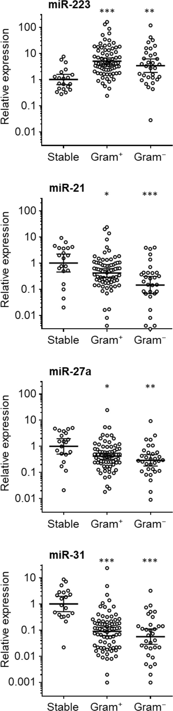
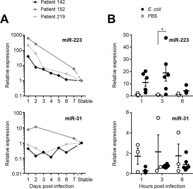
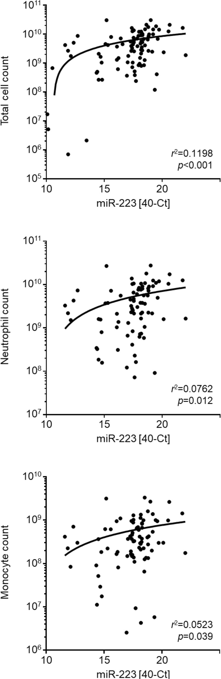
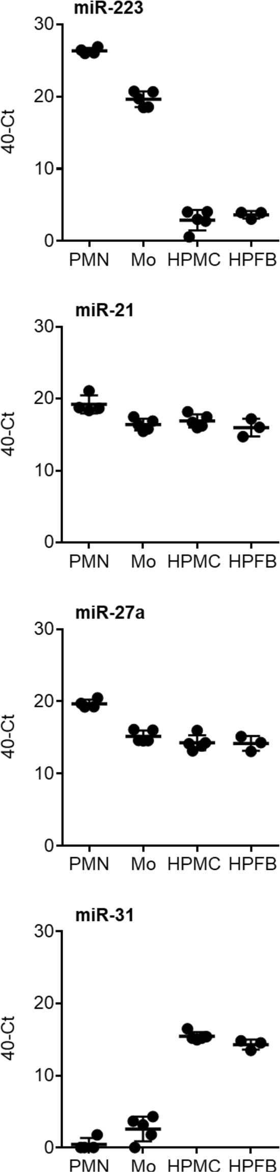
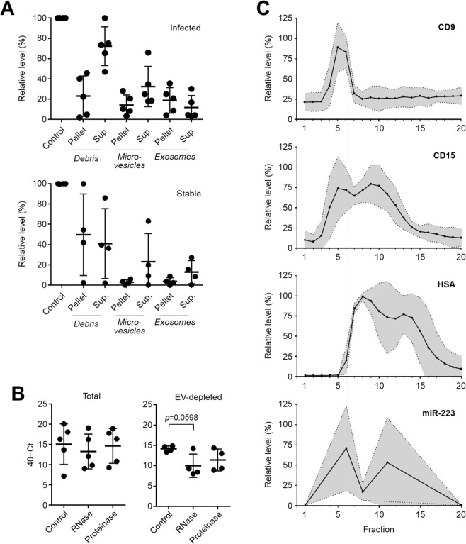
Infection remains a major cause of morbidity, mortality and technique failure in patients with end stage kidney failure who receive peritoneal dialysis (PD). Recent research suggests that the early inflammatory response at the site of infection carries diagnostically relevant information, suggesting that organ and pathogen-specific "immune fingerprints" may guide targeted treatment decisions and allow patient stratification and risk prediction at the point of care. Here, we recorded microRNA profiles in the PD effluent of patients presenting with symptoms of acute peritonitis and show that elevated peritoneal miR-223 and reduced miR-31 levels were useful predictors of bacterial infection. Cell culture experiments indicated that miR-223 was predominantly produced by infiltrating immune cells (neutrophils, monocytes), while miR-31 was mainly derived from the local tissue (mesothelial cells, fibroblasts). miR-223 was found to be functionally stabilised in PD effluent from peritonitis patients, with a proportion likely to be incorporated into neutrophil-derived exosomes. Our study demonstrates that microRNAs are useful biomarkers of bacterial infection in PD-related peritonitis and have the potential to contribute to disease-specific immune fingerprints. Exosome-encapsulated microRNAs may have a functional role in intercellular communication between immune cells responding to the infection and the local tissue, to help clear the infection, resolve the inflammation and restore homeostasis.


Conflict of interest statement

The authors declare no competing interests.

Figures

Figure 1
Figure 1
Peritoneal microRNAs in PD patients with and without acute bacterial peritonitis. Levels of miR-223, miR-21, miR-31 and miR-27a were measured in effluent samples from 20 stable PD patients, 76 patients with confirmed Gram+ infections and 31 with Gram infections, and normalised to snRNA U6. Each data point represents an individual patient; lines indicate geometric means and 95% confidence intervals. Data were analysed using Kruskal-Wallis tests combined with Dunn’s multiple comparisons tests versus stable controls.
Figure 2
Figure 2
Peritoneal microRNAs during bacterial peritonitis. (A) miR-223, miR-21, miR-27a and miR-31 were measured in effluent samples from three individual patients followed over the course of a week, starting from their presentation at hospital with acute peritonitis. All levels were normalised to snRNA U6 levels and are shown as relative values compared to a corresponding post-infection sample from each patient 10–12 months after the resolution of the peritonitis episode. Infective organisms were identified by microbiological culture as Staphylococcus aureus (#142), coagulase-negative Staphylococcus (#152) and alpha-haemolytic Streptococcus (#219). (B) microRNA expression during acute peritoneal E. coli infection in C57BL/6 mice aged 8–12 weeks (n = 6), compared to mock-treated animals (PBS; n = 3). Each data point represents an individual animal; lines indicate means and standard errors. Data were analysed by two-way ANOVA and Holm-Sidak’s multiple comparisons test.
Figure 3
Figure 3
Correlation of peritoneal microRNAs with cellular infiltration during peritonitis. Linear regression of peritoneal miR-223 levels (40−Ct) with cell numbers in the inflammatory infiltrate, shown as total cell count and the number of live CD14+ monocytes/macrophages or CD15+ neutrophils in effluent samples from 82 PD patients with acute peritonitis.
Figure 4
Figure 4
Cellular source of peritoneal microRNAs. Expression of microRNAs upon in vitro culture for 4 hours of neutrophils (PMN, n = 4) and monocytes (Mo, n = 5) from human blood and mesothelial cells (HPMC, n = 5) and fibroblasts (HPFB, n = 3) from human omentum. Each data point corresponds to an individual donor; lines indicate means and standard deviation.
Figure 5
Figure 5
Stabilisation of miR-223 in PD effluent by extracellular vesicles. (A) miR-223 levels in cell-free effluent from infected (top, n = 5) and stable (bottom, n = 4) PD patients, before (control) and after differential centrifugation to pellet cellular debris, larger microvesicles and smaller exosomes. Data are shown as mean values ± SD, in relation to the amount of miR-223 present in the unspun effluent (control) serving as reference. (B) Susceptibility of extracellular miR-223 in effluent from infected PD patients to RNase A and proteinase K treatment, before (left, n = 5) and after (right, n = 4) depletion of exosomes. Each data point corresponds to an individual patient, shown as raw 40−Ct values; lines indicate means and standard deviations. Statistical analysis was performed using Kruskal-Wallis tests combined with Dunn’s multiple comparisons tests. (C) Fractionation of cell-free effluent from five infected PD patients by size exclusion chromatography and detection of CD9, CD15 and human serum albumin (HSA) in each fraction using plate-bound immunoassays. miR-223 was only quantified in fractions 1, 6 (marked by the dashed line), 11 and 20. Graphs depict the relative levels of each marker compared to the fraction containing the maximum amount; lines show the means and the shaded areas the 95% confidence interval.

References

    1. Chakera A, et al. Peritonitis in peritoneal dialysis patients: the case for rapid diagnosis, targeted treatment, and monitoring to improve outcomes. EMJ. 2018;6:56–64.
    1. Szeto CC, et al. ISPD catheter-related infection recommendations: 2017 update. Perit. Dial. Int. 2017;37:141–154. doi: 10.3747/pdi.2016.00120. - DOI - PubMed
    1. Eberl M, Friberg IM, Liuzzi AR, Morgan MP, Topley N. Pathogen-specific immune fingerprints during acute infection: the diagnostic potential of human γδ T-cells. Front. Immunol. 2014;5:572. doi: 10.3389/fimmu.2014.00572. - DOI - PMC - PubMed
    1. Aufricht C, et al. Biomarker research to improve clinical outcomes of peritoneal dialysis: consensus of the European Training and Research in Peritoneal Dialysis (EuTRiPD) network. Kidney Int. 2017;92:824–835. doi: 10.1016/j.kint.2017.02.037. - DOI - PubMed
    1. Betjes MG, et al. Analysis of the peritoneal cellular immune system during CAPD shortly before a clinical peritonitis. Nephrol. Dial. Transplant. 1994;9:684–692. doi: 10.1093/ndt/9.6.684. - DOI - PubMed

Publication types

MeSH terms