. 2019 Sep 7;44(7):511-521.
doi: 10.1093/chemse/bjz046.

Differential Expression of Mucins in Murine Olfactory Versus Respiratory Epithelium

Affiliations

Differential Expression of Mucins in Murine Olfactory Versus Respiratory Epithelium

Christopher Kennel et al. Chem Senses. .

Abstract

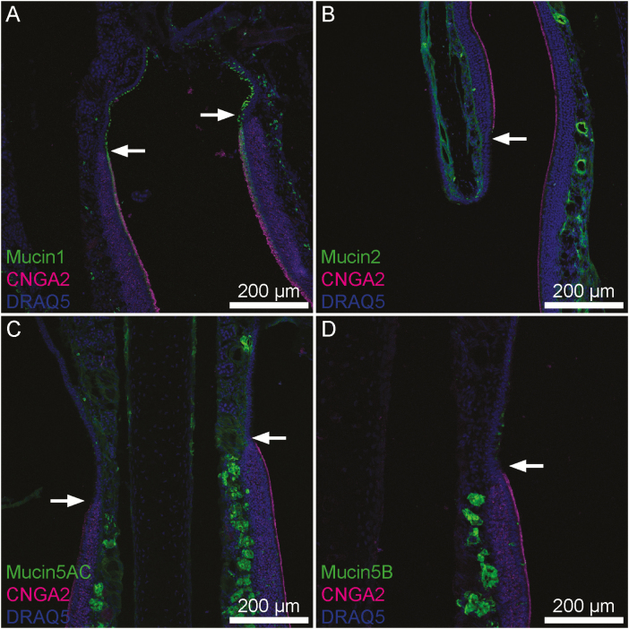
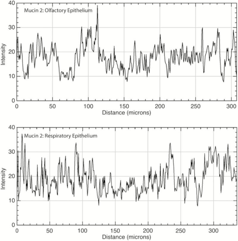
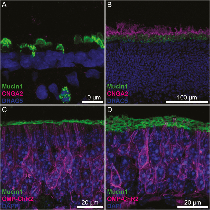
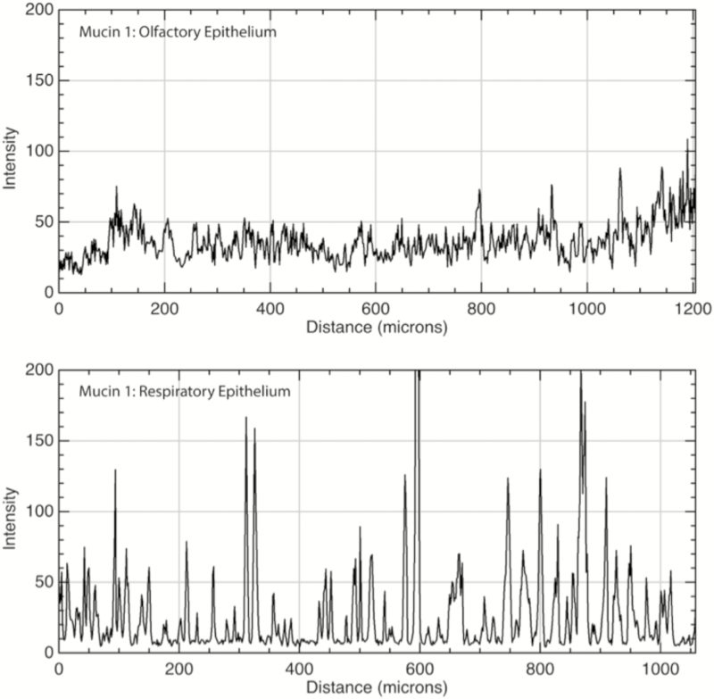
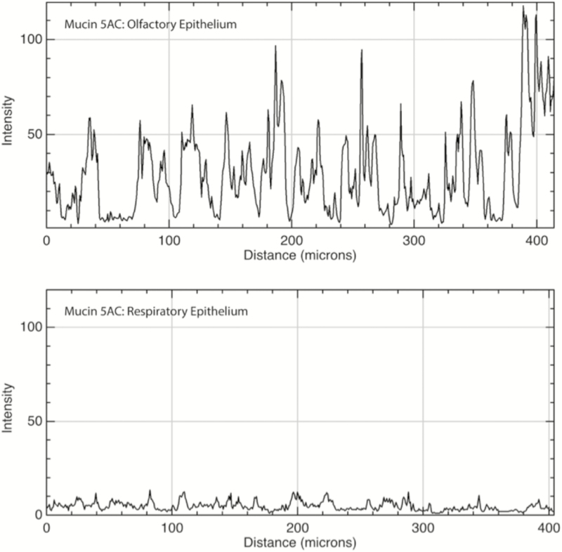
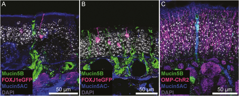
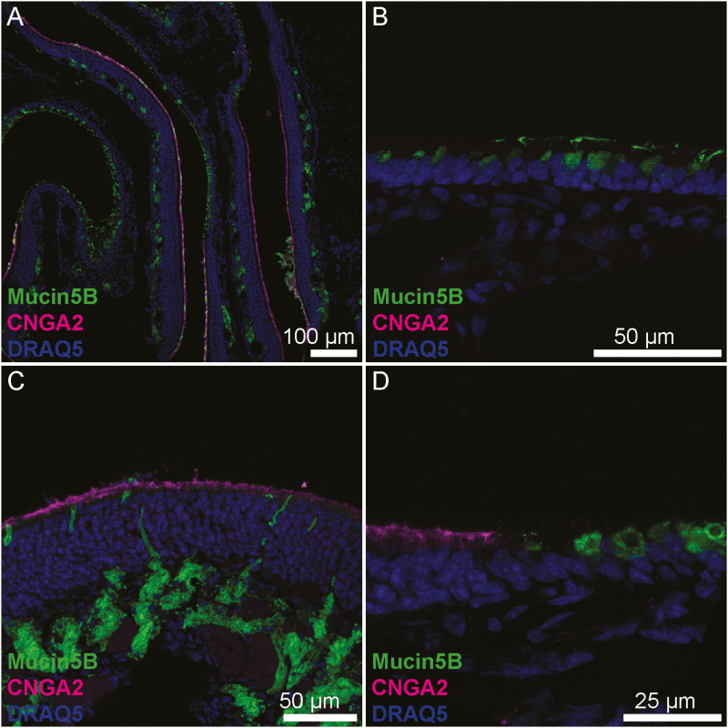
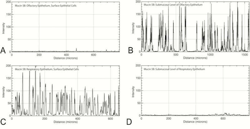
Mucins are a key component of the surface mucus overlying airway epithelium. Given the different functions of the olfactory and respiratory epithelia, we hypothesized that mucins would be differentially expressed between these 2 areas. Secondarily, we evaluated for potential changes in mucin expression with radiation exposure, given the clinical observations of nasal dryness, altered mucus rheology, and smell loss in radiated patients. Immunofluorescence staining was performed to evaluate expression of mucins 1, 2, 5AC, and 5B in nasal respiratory and olfactory epithelia of control mice and 1 week after exposure to 8 Gy of radiation. Mucins 1, 5AC, and 5B exhibited differential expression patterns between olfactory and respiratory epithelium (RE) while mucin 2 showed no difference. In the olfactory epithelium (OE), mucin 1 was located in a lattice-like pattern around gaps corresponding to dendritic knobs of olfactory sensory neurons, whereas in RE it was intermittently expressed by surface goblet cells. Mucin 5AC was expressed by subepithelial glands in both epithelial types but to a higher degree in the OE. Mucin 5B was expressed by submucosal glands in OE and by surface epithelial cells in RE. At 1-week after exposure to single-dose 8 Gy of radiation, no qualitative effects were seen on mucin expression. Our findings demonstrate that murine OE and RE express mucins differently, and characteristic patterns of mucins 1, 5AC, and 5B can be used to define the underlying epithelium. Radiation (8 Gy) does not appear to affect mucin expression at 1 week.

Level of evidence: N/A (Basic Science Research).IACUC-approved study [Protocol 200065].

Keywords: MUC1; MUC5; mucin; olfaction; radiation; sinus.


Figures

Figure 1.
Figure 1.
Transitions between OE and RE in mouse nasal tissue. CNGA2 (magenta), a marker specific to neurocilia of OSNs indicates portions of OE. Visible portions of OE and RE are broadly representative of these tissues. (A) Mucin 1 (green) is present uniformly across a continuous layer of sustentacular cells within the OE whereas in the RE scattered individual surface epithelial cells express it. (B) Mucin 2 (green) is expressed uniformly within lamina propria connective tissue below both OE and RE. (C) Mucin 5AC is expressed by submucosal glands and to a lesser degree within lamina propria connective tissue below both OE and RE. The density of positively staining glands was much higher in the OE. (D) Mucin 5B was expressed intensely by submucosal Bowman’s glands within the OE, but within the RE goblet cells expressed mucin 5B. Arrows denote the transition from RE to OE. Also, this transition is noticeable as the thickness of the epithelial layer changes from thick to thin from OE to RE.
Figure 2.
Figure 2.
Immunofluorescence quantitation of mucin 2 in the lamina propria underlying OE and RE. Mucin 2 was diffusely present within the lamina propria of both the olfactory (top panel) and respiratory (bottom panel) epithelia. X axis is the distance moving along a linear region of interest within the lamina propria, parallel to the epithelial layer; Y axis is intensity of immunofluorescence with possible values ranging from 0 to 255. Mean OE versus RE immunofluorescence was 19.0 versus 17.7, respectively (P < 0.01).
Figure 3.
Figure 3.
Mucin 1 differential staining between OE and RE. (A) Mucin 1 is present in the apical layer of RE and expressed by scattered individual cells. (B and C) In the OE, mucin 1 lies at the base of the neurocilia. When viewed in oblique section (B), mucin 1 exhibits a lattice-like staining pattern indicative of perforations by OSN dendritic knobs suggesting it may be produced and secreted into this layer by the sustentacular cells. (C and D) OMP-ChR2-YFP knock-in mice were used to examine the relationship between OSNs (magenta) and mucin 1 (green). Mucin 1 is found in a layer just above the sustentacular cells, at the level of the OSN dendritic knob, as olfactory dendrites coursing through the OE are seen. In orthogonal section, this layer appears continuous, but in an oblique section (D) the lattice pattern of mucin 1 is clearly observed.
Figure 4.
Figure 4.
Immunofluorescence quantitation of mucin 1 in OE and RE. Top: Mucin 1 was diffusely present along the apical surface of olfactory epithelial cells. Bottom: In the RE, it was present on the apical surfaces of individual goblet cells, resulting in a series of immunofluorescence peaks. X axis is distance along a linear region of interest consisting of the apical surface of the epithelium; Y axis is intensity of immunofluorescence with possible values ranging from 0 to 255. Values indicate consistent moderate expression of the membrane-bound protein throughout the OE, versus intermittent areas of high expression in RE consistent with goblet cell colocalization.
Figure 5.
Figure 5.
Immunofluorescence quantitation of mucin 5AC in OE and RE. Top: Mucin 5AC was present primarily in submucosal glands within the OE and to a much lesser degree within the surrounding lamina propria connective tissue. Bottom: In the RE, there was a low level of immunofluorescence within the connective tissue and very few glands expressing the protein (none are present in the sample mentioned earlier). X axis is distance along a linear region of interest consisting of the submucosa, parallel to the epithelium; Y axis is intensity of immunofluorescence with possible values ranging from 0 to 255.
Figure 6.
Figure 6.
Mucin 5AC and 5B staining in OE. (A and B) FOXJ1-eGFP mice were used to visualize a small subset of OSNs. The cilia and dendritic knob of an OSN (magenta) are observed within a layer of mucin 5AC immunoreactivity (blue; A), though this layer was not consistent across the entire OE (B). Additionally, mucin 5B+ (green), AC—Bowman’s glands are present in the OE. (C) Some Bowman’s glands were immunoreactive for mucin 5AC, surrounded by OMP-ChR2+ OSNs.
Figure 7.
Figure 7.
Mucin 5B differential staining between OE and RE. (A) In the OE, mucin 5B is located in submucosal glands while in the RE it is secreted by surface goblet cells. (B) Magnification of RE showing goblet cells secreting directly to the epithelial surface. Note the vesicles filled with mucin oriented to the apical side while the nuclei are located basolaterally. (C) Magnification of OE showing submucosal glands with ducts to the luminal surface. (D) Transition between OE (left) and RE (right).
Figure 8.
Figure 8.
Immunofluorescence quantitation of mucin 5B at the epithelial and submucosal level of OE and RE. Mucin 5B exhibited almost no immunofluorescence in the epithelial layer (A), but was present primarily in submucosal glands of the OE as evidenced by the strong peaks of immunofluorescence (B). Individual goblet cells throughout the surface of the RE expressed high levels of mucin 5B, resulting in immunofluorescence peaks (C). There was virtually no mucin 5B in the submucosal level of RE (D). X axis is distance along a linear region of interest; Y axis is intensity of immunofluorescence with possible values ranging from 0 to 255.

Similar articles

Cited by

References

    1. Abramoff M, Magalhaes P, Ram S. 2004. Image processing with ImageJ. Biophotonics Int. 11(7):36–42.
    1. Ali MS, Pearson JP. 2007. Upper airway mucin gene expression: a review. Laryngoscope. 117:932–938. - PubMed
    1. Ali MS, Wilson JA, Bennett M, Pearson JP. 2005. Mucin gene expression in nasal polyps. Acta Otolaryngol. 125:618–624. - PubMed
    1. Ali MS, Wilson JA, Pearson JP. 2002. Mixed nasal mucus as a model for sinus mucin gene expression studies. Laryngoscope. 112:326–331. - PubMed
    1. Axel R. 2005. Scents and sensibility: a molecular logic of olfactory perception (Nobel lecture). Angew Chem Int Ed Engl. 44(38):6110–6127. - PubMed

Publication types