

DeepHarmony: A deep learning approach to contrast harmonization across scanner changes
- PMID: 31301354
- PMCID: PMC6874910
- DOI: 10.1016/j.mri.2019.05.041
DeepHarmony: A deep learning approach to contrast harmonization across scanner changes
Abstract
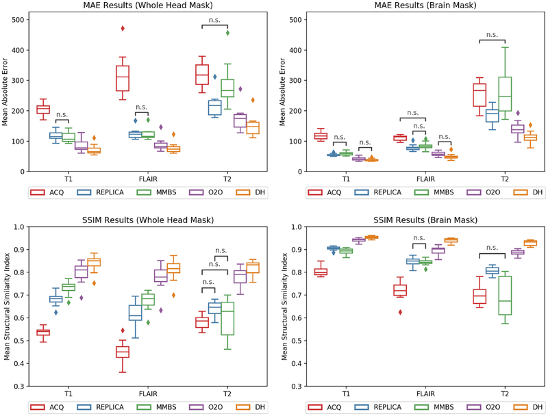
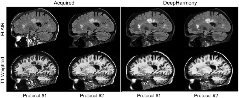
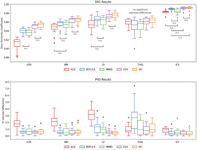
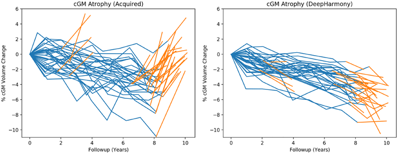
Magnetic resonance imaging (MRI) is a flexible medical imaging modality that often lacks reproducibility between protocols and scanners. It has been shown that even when care is taken to standardize acquisitions, any changes in hardware, software, or protocol design can lead to differences in quantitative results. This greatly impacts the quantitative utility of MRI in multi-site or long-term studies, where consistency is often valued over image quality. We propose a method of contrast harmonization, called DeepHarmony, which uses a U-Net-based deep learning architecture to produce images with consistent contrast. To provide training data, a small overlap cohort (n = 8) was scanned using two different protocols. Images harmonized with DeepHarmony showed significant improvement in consistency of volume quantification between scanning protocols. A longitudinal MRI dataset of patients with multiple sclerosis was also used to evaluate the effect of a protocol change on atrophy calculations in a clinical research setting. The results show that atrophy calculations were substantially and significantly affected by protocol change, whereas such changes have a less significant effect and substantially reduced overall difference when using DeepHarmony. This establishes that DeepHarmony can be used with an overlap cohort to reduce inconsistencies in segmentation caused by changes in scanner protocol, allowing for modernization of hardware and protocol design in long-term studies without invalidating previously acquired data.
Keywords: Contrast harmonization; Deep learning; Magnetic resonance imaging.
Copyright © 2019 Elsevier Inc. All rights reserved.
Conflict of interest statement
Declaration of Interests:
Blake E. Dewey: Mr. Dewey is partially supported by a grant from Biogen.
Can Zhao: Ms. Zhao is partially supported by a grant from 12Sigma. Jacob C. Reinhold: Mr. Reinhold is partially supported by a grant from 12Sigma.
Aaron Carass: Mr. Carass is partially supported by a grant from 12Sigma and Biogen.
Kathryn C. Fitzgerald: Nothing to declare.
Elias S. Sotirchos: Nothing to declare.
Shiv Saidha: Dr. Saidha has received consulting fees from Medical Logix and Axon Advisors LLC, speaking honoraria from the National Association of Managed Care Physicians, Family Medicine Foundation of West Virginia, and Advanced Studies in Medicine and served on scientific advisory boards for Biogen-Idec, Genzyme, Genentech Corporation & Novartis. He receives research support from Genentech Corporation and the National MS Society, and received support from the Race to Erase MS foundation.
Jiwon Oh: Nothing to declare.
Dzung L. Pham: Nothing to declare.
Peter Calabresi: Dr. Calabresi has received personal consulting fees from Biogen. Dr. Calabresi is PI on grants to JHU from MedImmune, Annexon, Teva, Novartis, Genzyme and Biogen.
Peter C. M. van Zijl: Dr. van Zijl is a paid lecturer for Philips Medical Systems.
Jerry L. Prince: Dr. Prince is PI on grants to JHU from Biogen and 12Sigma.
Figures
References
-
- Shinohara RT, Oh J, Nair G, Calabresi PA, Davatzikos C, Doshi J,Henry RG, Kim G, Linn KA, Papinutto N, Pelletier D, Pham DL,Reich DS, Rooney W, Roy S, Stern W, Tummala S, Yousuf F,Zhu A, Sicotte NL, Bakshi R, NAIMS Cooperative, Volumetric Analysis from a Harmonized Multisite Brain MRI Study of a Single Subject with Multiple Sclerosis., American Journal of Neuroradiology 38 (8) (2017) 1501–1509, doi: 10.3174/ajnr.A5254. - DOI - PMC - PubMed
-
- Han X, Jovicich J, Salat D, van der Kouwe A, Quinn B, Czanner S,Busa E, Pacheco J, Albert M, Killiany R, Maguire P, Rosas D,Makris N, Dale A, Dickerson B, Fischl B, Reliability of MRI-derived measurements of human cerebral cortical thickness: The effects of field strength, scanner upgrade and manufacturer, NeuroImage 32 (1) (2006) 180–194, ISSN 1053–8119, doi: 10.1016/j.neuroimage.2006.02.051. - DOI - PubMed
-
- Biberacher V, Schmidt P, Keshavan A, Boucard CC, Righart R,Smann P, Preibisch C, Frbel D, Aly L, Hemmer B, Zimmer C, Henry RG, Mhlau M, Intra- and interscanner variability of magnetic resonance imaging based volumetry in multiple sclerosis, NeuroImage 142 (2016) 188–197, ISSN 1053–8119, doi: 10.1016/j.neuroimage.2016.07.035. - DOI - PubMed
-
- Schnack HG, van Haren NE, Brouwer RM, van Baal GCM,Picchioni M, Weisbrod M, Sauer H, Cannon TD, Huttunen M,Lepage C, Collins DL, Evans A, Murray RM, Kahn RS, Hulsho Pol HE, Mapping reliability in multicenter MRI: Voxel-based morphometry and cortical thickness, Human Brain Mapping 31 (12) (2010) 1967–1982, doi: 10.1002/hbm.20991. - DOI - PMC - PubMed
Publication types
MeSH terms
Grants and funding
LinkOut - more resources
Full Text Sources
Other Literature Sources
Medical
