Review
. 2019 Nov:73:100769.
doi: 10.1016/j.preteyeres.2019.07.002. Epub 2019 Jul 10.

Axon injury signaling and compartmentalized injury response in glaucoma

Affiliations
Review

Axon injury signaling and compartmentalized injury response in glaucoma

Stephanie B Syc-Mazurek et al. Prog Retin Eye Res. 2019 Nov.

Abstract

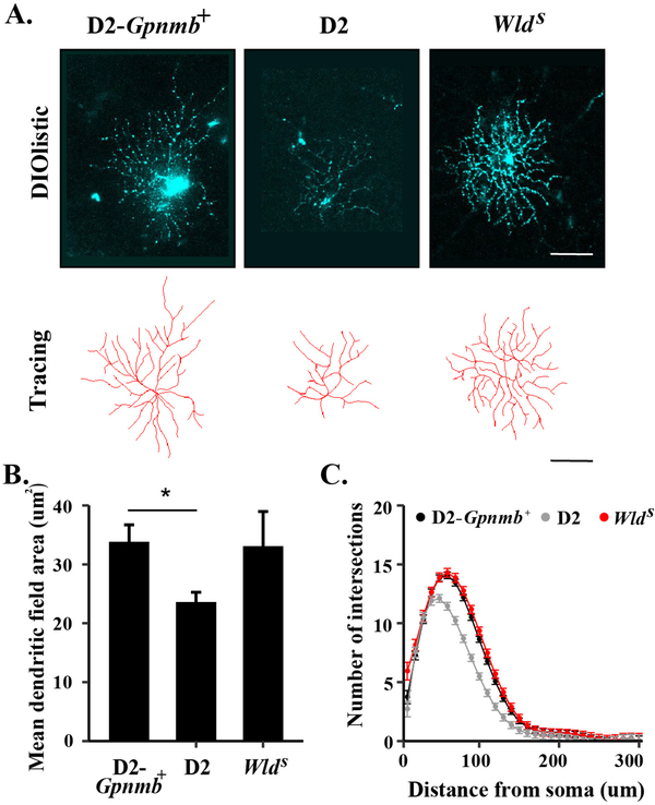
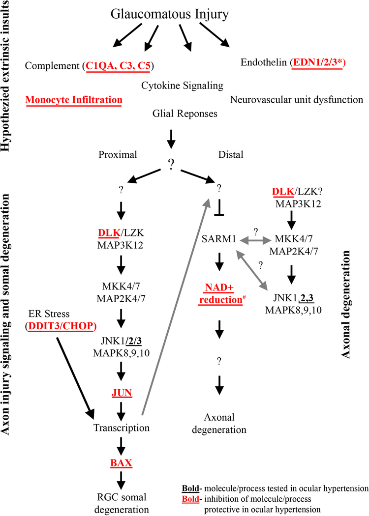
Axonal degeneration is an active, highly controlled process that contributes to beneficial processes, such as developmental pruning, but also to neurodegeneration. In glaucoma, ocular hypertension leads to vision loss by killing the output neurons of the retina, the retinal ganglion cells (RGCs). Multiple processes have been proposed to contribute to and/or mediate axonal injury in glaucoma, including: neuroinflammation, loss of neurotrophic factors, dysregulation of the neurovascular unit, and disruption of the axonal cytoskeleton. While the inciting injury to RGCs in glaucoma is complex and potentially heterogeneous, axonal injury is ultimately thought to be the key insult that drives glaucomatous neurodegeneration. Glaucomatous neurodegeneration is a complex process, with multiple molecular signals contributing to RGC somal loss and axonal degeneration. Furthermore, the propagation of the axonal injury signal is complex, with injury triggering programs of degeneration in both the somal and axonal compartment. Further complicating this process is the involvement of multiple cell types that are known to participate in the process of axonal and neuronal degeneration after glaucomatous injury. Here, we review the axonal signaling that occurs after injury and the molecular signaling programs currently known to be important for somal and axonal degeneration after glaucoma-relevant axonal injuries.

Keywords: Apoptosis; Axon; Axonopathy; Dendritic remodeling; Intraocular pressure; Neuroprotection; Optic neuropathy; Synaptic loss.


Figures

Figure 1:
Figure 1:. BAX deficiency protects RGC somas from glaucomatous damage and the WldS allele protects both somas and axons
BAX deficiency protects RGC somas in the DBA/2J model of ocular hypertension. The corresponding retinas of severely glaucomatous optic nerves (95% or more axonal degeneration) from BAX deficient mice (Bax−/−) had significantly more RGCs as compared to wildtype animals (Bax+/+, A and B, Nissel-stained with cresyl violet, Scale bar = 50 μm). BAX deficient animals have significantly increased RGCs at baseline (BAX is required for normal developmental RGC apoptosis) and thus quantification is presented as percent surviving RGCs (retinas from corresponding optic nerves with severely glaucomatous retinas as compared to retinas with corresponding optic nerves with no glaucomatous damage from the same genotype, E). Optic nerve cross-sections stained with p-phenylenediamine (PPD) demonstrate glial scarring and loss of axons in both wildtype and BAX deficient animals (G and H, Scale bar = 50 μm). BAX deficiency does not prevent glaucomatous axonal degeneration (K). The WldS allele protects both axons and somas from glaucomatous damage. By rescuing approximately 50% of optic nerves from severe glaucoma, the WldS allele also protected RGCs in the corresponding retinas of these nerves (C- severe glaucoma wildtype DBA/2J and D- no/early glaucoma with WldS allele, Scale bar = 100 μm). In retinas with corresponding severe nerves, the WldS allele did not protect RGCs somas (F). As compared with wildtype animals, optic nerves of animals carrying the WldS allele (hemi) had less glaucomatous damage as compared to wildtype animals. Optic nerve cross-sections stained with PPD demonstrate glial scarring and loss of axons in the wildtype glaucomatous DBA/2J nerves but not in those carrying the WldS allele (I and J, Scale bar = 100 μm). BAX heterozygotes (Bax+/−) were not significantly different from wildtype DBA/2J animals (L, Figure adapted from Howell et al., 2007; Libby et al., 2005b).
Figure 2:
Figure 2:. Compartmentalization of axonal degeneration and somal apoptosis
Schematic of axonal injury leading to both somal apoptosis and axonal degeneration. Inciting extrinsic factors lead to injury at the glial lamina after ocular hypertensive injury. Axonal injury signaling in the proximal and distal axon leads to degeneration of both somal and axonal compartment. Somal degeneration is known to occur through transcription and subsequent BAX-dependent apoptosis while the WldS allele is known to protect the axonal compartment from degeneration. Furthermore, anterograde signaling from the somal compartment contributes to axonal degeneration suggesting that while axonal injury is a major driver of pro-degenerative signals, there are interactions between the two compartments (Simon et al., 2016).
Figure 3:
Figure 3:. Somal apoptotic signaling in glaucomatous neurodegeneration
Diagram of the molecules that contribute to somal degeneration in glaucoma-relevant axonal injury. The mitogen activated protein kinase family and endoplasmic reticulum are important for pro-apoptotic signaling in glaucomatous neurodegeneration. MAPK3 kinases, DLK and LZK, activate the MAP2Ks, MKK4 and MKK7, which in turn activate the MAPKs, JNK1–3 and their canonical target JUN. The transcription factors JUN and DDIT3/CHOP lead to transcriptional changes that ultimate culminate in BAX activation and RGC apoptosis (Figure adapted from Fernandes et al., 2018).
Figure 4:
Figure 4:. Somal apoptosis is controlled by JUN and DDIT (CHOP)
The majority of somal apopotic signaling in RGCs is controlled by the transcription factors JUN, a member of MAPK signaling, and DDIT (CHOP), a key mediator of endoplasmic reticulum stress. Animals deficient in both of these molecules had sustained robust protection after optic nerve crush injury (CONC) as compared to animals individually deficient in JUN or DDIT. Representative retinal flat mount images with TUJ-1 staining, a marker of RGCs, (A) and quantification of TUJ-1+ cells (B) are presented. RGC survival significantly differed (P<0.01) between DDIT3, JUN, and dual DDIT3/JUN deficient animals at all time points with the exception of between JUN and dual DDIT3/JUN at the 14 day time point which was nonsignificant. Data are presented as the percent of RGCs surviving in the CONC animals relative to sham animals (Scale bar = 50 um, Figure from Syc-Mazurek et al., 2017b).
Figure 5:
Figure 5:. Axonal degeneration signaling in glaucomatous neurodegeneration
Schematic of the molecules thought to contribute to glaucomatous neurodegeneration. Multiple possible mechanisms are thought to lead to decreased levels of NMNAT after injury. Axonal injury interrupts the anterograde transport of cytoplasmic NMNAT2 from the cell body and activation of JNK signaling leads to NMNAT2 turnover. The axonal protection afforded by animals carrying the WldS allele is thought to occur through NMNAT1 substituting for NMNAT2. Decreased levels of NMNAT2 can lead to accumulation of nicotinamide (NMN) which has been shown to be pro-degenerative at certain levels in other systems (Di Stefano et al., 2015). Nicotinamide is also a precursor of NAD+ and oral administration of nicotinamide or increasing expression of Nmnat1 (an enzyme that produces NAD+) in ocular hypertensive animals is protective from glaucomatous damage (Williams et al., 2017b). Decreased NMNAT2 activity subsequently leads to activation of SARM NADase activity likely via an increase in intracellular calcium, and additional work will be needed to determine the exact sequence of events in glaucomatous neurodegeneration given the potential complexity of NMN signaling. JNK can also activate SARM NADase activity via phosphorylation of SARM. Ultimately these events lead to deceased NAD+ levels leading to decreased ATP (which can also occur through SARM-JNK signaling), calpain activation and axon degradation. Reprinted from Biochemical Pharmacology, 161 (2019), Michael Carty and Andrew G. Bowie, SARM: from immune regulator to cell executioner, 52–62, 2019 with permission from Elsevier (Carty and Bowie, 2019).
Figure 6:
Figure 6:. The WldS allele prevents ocular hypertensive induced dendritic remodeling
Retinal cells were labeled using DiO/Dil bullets in 9 month D2-Gpnmb+, wildtype DBA/2J (D2), and DBA/2J animals carrying the WldS allele and RGCs were subsequently selected for analysis (A). While there was a significant decrease (*) in mean dendritic field area in RGCs from wildtype D2 animals as compared to D2-Gpnmb+ animals, there was no difference between mean field area (B) or number of intersections (C, as determined by Sholl analysis) between RGCs from D2-Gpnmb+ animals and those DBA/2J animals carrying the WldS allele. These results demonstrate that presence of the WldS allele prevents ocular hypertension induced dendritic changes (Figure from Harder et al., 2017).
Figure 7:
Figure 7:. Molecular program of somal apoptosis and axonal degeneration after axonal injury
Diagram of the molecules that contribute to somal and axonal degeneration in glaucoma-relevant axonal injury. Molecules in bold have been tested in ocular hypertension and deficiency of the molecules/processes in bold with red font have been shown to be protective for somal apoptosis in ocular hypertension. Question marks indicate areas currently unknown or areas where research has shown potential for bidirectional signaling. Multiple extrinsic events are thought to be important for early changes in glaucomatous neurodegeneration, but the ordering of these early changes and the possible interactions between these early changes is still poorly understood. * The role of endothelin was tested using a pan endothelin antagonist (Howell et al., 2011a). # Inhibition of the reduction of NAD+ has been tested in models of ocular hypertension with oral administration of nicotinamide, an NAD+ precursor, and using gene therapy to increase expression of Nmnat1 (Williams et al., 2017b). Anterograde transcriptional changes from the somal compartment are also thought to influence axonal degeneration signaling (large gray arrow, components of figure adapted from Fernandes et al., 2018).

References

    1. Agostinone J, Di Polo A, 2015. Retinal ganglion cell dendrite pathology and synapse loss: Implications for glaucoma. Prog Brain Res 220, 199–216. - PubMed
    1. Akhter R, Sanphui P, Das H, Saha P, Biswas SC, 2015. The regulation of p53 up-regulated modulator of apoptosis by JNK/c-Jun pathway in beta-amyloid-induced neuron death. J Neurochem 134, 1091–1103. - PubMed
    1. Ali YO, Li-Kroeger D, Bellen HJ, Zhai RG, Lu HC, 2013. NMNATs, evolutionarily conserved neuronal maintenance factors. Trends Neurosci 36, 632–640. - PMC - PubMed
    1. Almasieh M, Wilson AM, Morquette B, Cueva Vargas JL, Di Polo A, 2012. The molecular basis of retinal ganglion cell death in glaucoma. Prog Retin Eye Res 31, 152–181. - PubMed
    1. Ambacher KK, Pitzul KB, Karajgikar M, Hamilton A, Ferguson SS, Cregan SP, 2012. The JNK- and AKT/GSK3beta- signaling pathways converge to regulate Puma induction and neuronal apoptosis induced by trophic factor deprivation. PLoS One 7, e46885. - PMC - PubMed

Publication types