. 2019 Sep:27:47-61.
doi: 10.1016/j.molmet.2019.06.023. Epub 2019 Jul 1.

Evaporative cooling provides a major metabolic energy sink

Affiliations

Evaporative cooling provides a major metabolic energy sink

Ildiko Kasza et al. Mol Metab. 2019 Sep.

Abstract

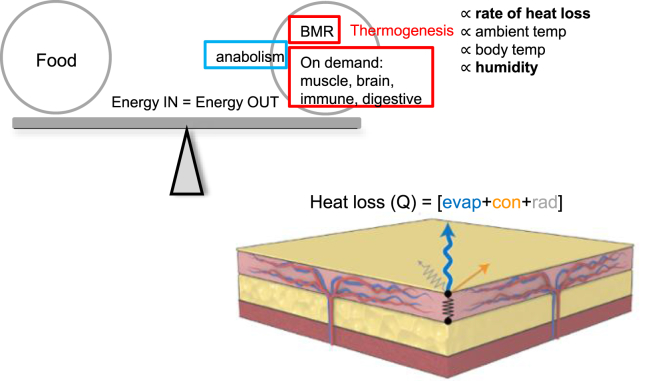
Objective: Elimination of food calories as heat could help redress the excess accumulation of metabolic energy exhibited as obesity. Prior studies have focused on the induction of thermogenesis in beige and brown adipose tissues as the application of this principle, particularly because the β-adrenergic environment associated with thermogenic activation has been shown to have positive health implications. The counterpoint to this strategy is the regulation of heat loss; we propose that mammals with inefficient heat conservation will require more thermogenesis to maintain body temperature.

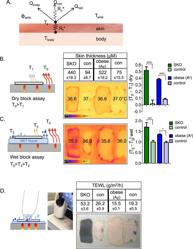
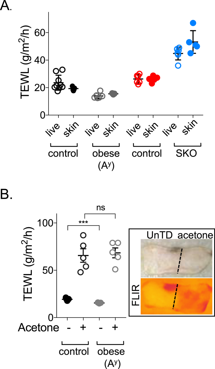
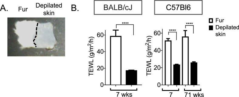
Methods: Surface temperature thermography and rates of trans-epidermal water loss were integrated to profile the total heat transfer of genetically-engineered and genetically variable mice.

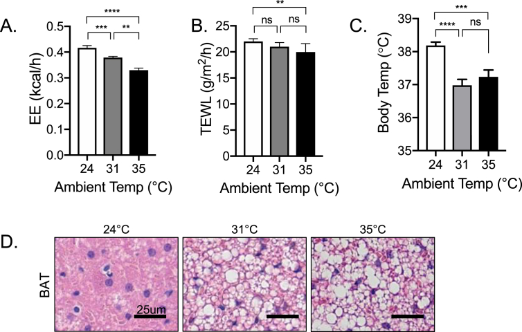
Results: These data were incorporated with energy expenditure data to generate a biophysical profile to test the significance of increased rates of evaporative cooling.

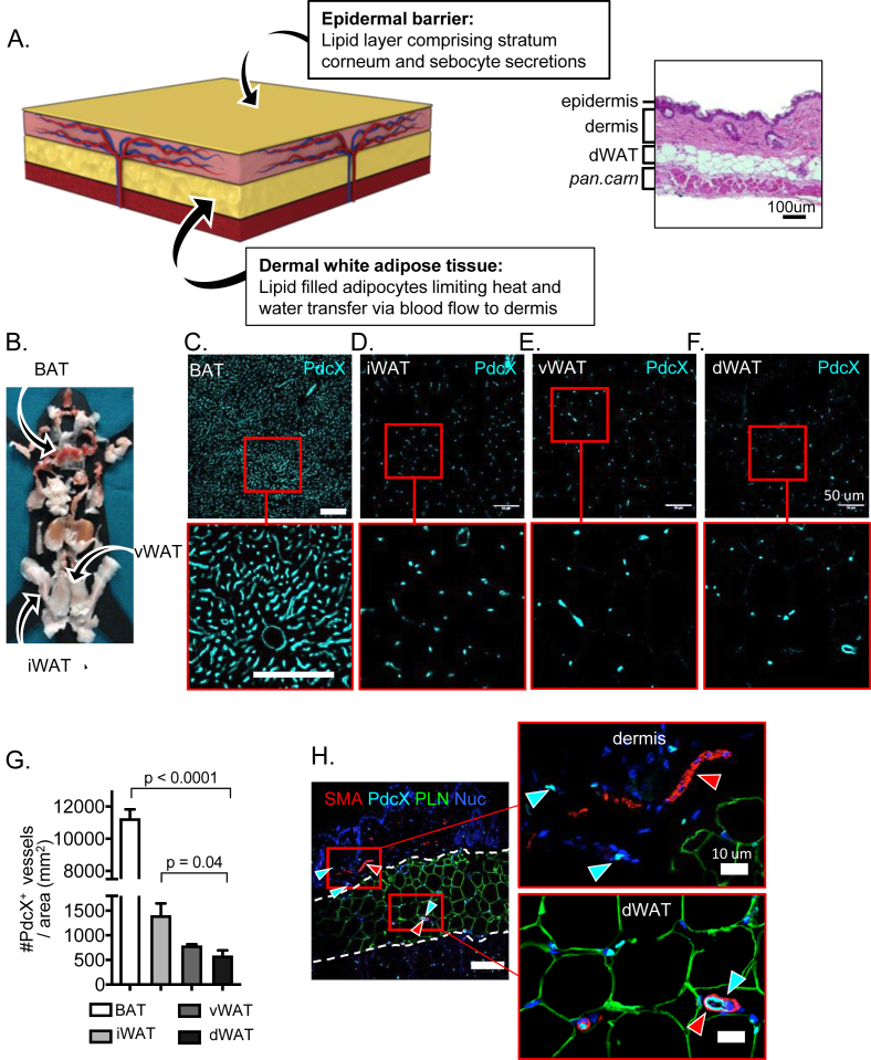
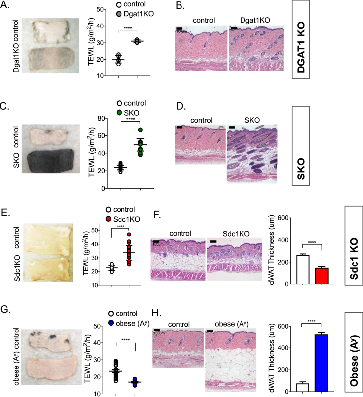
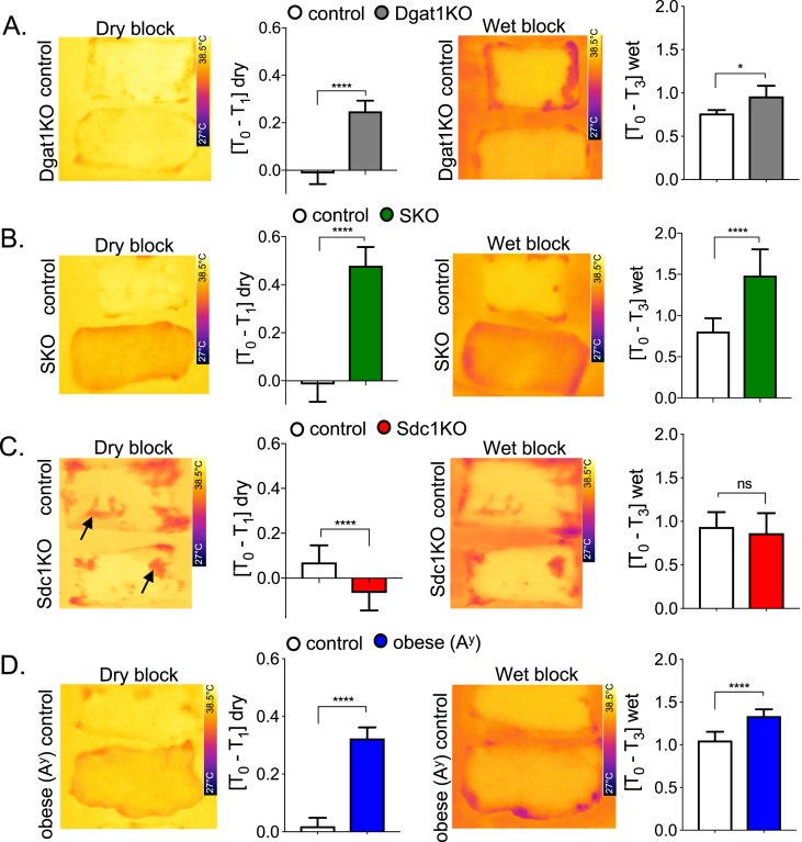
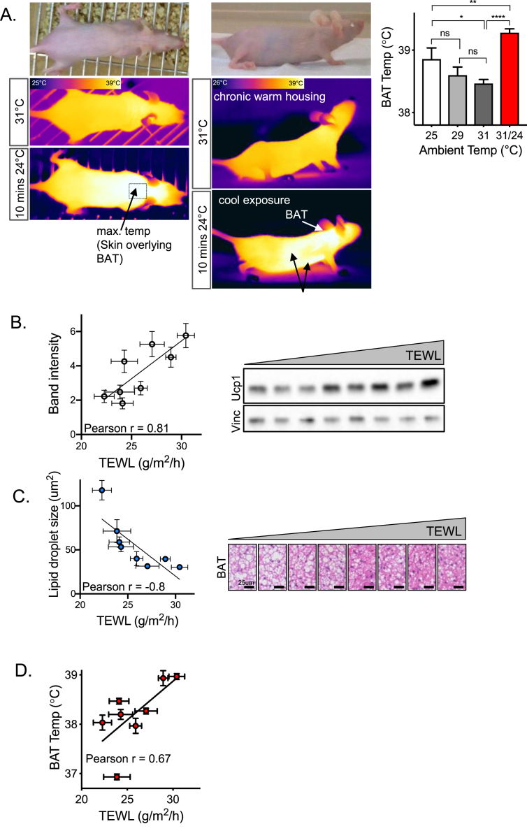
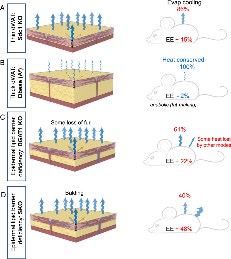
Conclusions: We show that mouse skins vary considerably in their heat retention properties, whether because of naturally occurring variation (SKH-1 mice), or genetic modification of the heat-retaining lipid lamellae (SCD1, DGAT1 or Agouti Ay obese mice). In particular, we turn attention to widely different rates of evaporative cooling as the result of trans-epidermal water loss; higher rates of heat loss by evaporative cooling leads to increased demand for thermogenesis. We speculate that this physiology could be harnessed to create an energy sink to assist with strategies aimed at treating metabolic diseases.

Keywords: Brown adipose tissue; Dermal white adipose tissue; Energy expenditure; Epidermal barrier; Evaporative cooling; Mouse skin; Obesity; Syndecan-1; Thermogenesis; Trans-epidermal water loss.


Figures

Figure 1
Figure 1
The structure of mouse skin. A. A scheme of skin structure illustrates two lipid bio-membranes, colored in yellow. The outer bio-membrane of the ceramide-enriched epidermis is the water-proofing component of skin: ceramide-based lipids of the stratum corneum generate a highly ordered gel phase membrane with restricted mobility, called the epidermal barrier . Junctions between keratinocytes and regulated water flow through aquaporins comprise a key “active” component of the epidermal barrier, reactive on acute time scales; on a slower time scale (days), the lipid composition of the stratum corneum changes as the superficial layers are sloughed and renewed . The thickness of the dermal white adipose tissue (dWAT) is regulated by interaction with microbes, ambient temperature and the stage of the hair cycle, and by genetics, diet and sex , . In between lies the dermis, laced with two vascular beds to allow adjustable blood volumes to control heat loss. At the right, for comparison, an H&E stained cross section of mouse skin (panniculus carnosus muscle layer; pan carn). Four adipose depots with different functions were collected for evaluation of vascular density (sites of each depot shown on left; B); dWAT, inguinal (“subcutaneous”) white adipose tissue (iWAT), brown adipose tissue (BAT) and visceral white adipose tissue (vWAT). Sections of each type were stained for podocalyxin, a vascular-associated antigen (C–F), and the density of vessels quantified (G). Sections of each were also stained with smooth muscle actin (SMA) to reveal pericyte-supported blood vessels, perilipin (PLN) to outline adipocytes and DAPI (Nuc) to counter-stain nuclei (H). The scale bar shown is 50 μM and applies to all panels C-F.
Figure 2
Figure 2
Illustration of the impact of evaporative cooling on heat transfer. A. Scheme of potential modes of heat loss. Loss of heat from body temperature (Tbody) to the outside (Tamb) is governed by at least three mechanisms that relate to the resistance of the skin biofabric (described by a factor, Rf) and the biophysical properties of the boundary layer (described by another factor, Rc). Heat can be lost by conduction (Qcon), radiation (Qrad) or evaporation (Qevap); together, these determine the surface temperature (Ts). The rate of radiation is controlled by the gradient of temperature from body to environment (Tamb); conduction by contact surfaces, and evaporative cooling by ambient humidity (ɸamb). B. Dry block assay. Skins were excised from mice, transferred to a heat block (37oC), and promptly imaged using infrared thermography (FLIR). Skin thickness was measured by histological analysis of sections. Impeded heat flow was visualized as a lower surface temperature (color scale shown at foot of image), and quantified as [T0-T1]dry, where higher values show higher insulation. C. Wet block assay. Skins were transferred to a water-saturated tissue substrate (emissivity >0.98, homogeneous background; 37 °C), and imaged by FLIR. The surface temperature recorded is lower for skins with high rates of transpiration and also lower for skins with more insulation (opposing metabolic effects). The surface temperature is quantified as [T0-T3]wet, where values combine these two distinct aspects of skin physiology. D. Trans-epidermal water loss assay reveals basis for discrepant dry and wet block images. TEWL was measured using a closed-chamber instrument; results for these skins varied over 3-fold, from skins ranging in thickness by nearly 6-fold (75 and 522 μM). Note that SKO skins appear black due to trapped hair follicles (see detailed descriptions in legend to Figure 4). Thermographic images were analyzed using the FLIR camera software as per Materials and Methods section; shown are mean values ± SEM, analyzed by unpaired 2-tailed t tests (****P < 0.0001; ***P < 0.001; *P < 0.05).
Figure 3
Figure 3
Calibrating the TEWL assay. A. Trans-epidermal water loss was compared for the skin of live mice, versus their pelts after euthanasia, for two types of mice included in this study and their respective controls, obese (Ay) and SKO (SCD1 skin specific knockout). B. Skins of control and obese mice were wiped with acetone to extract the lipid from the epidermal barrier, by way of positive control for the TEWL assay. Increased evaporative cooling is also visualized as a lower surface temperature assessed by infrared thermography for control mice (FLIR; right hand side). The acetone extraction protocol does not affect dWAT (confirmed by histological analysis of skins after treatment, data not shown). Data are expressed as mean ± SEM. Statistical analysis was performed with unpaired 2-tailed t test (***P < 0.001, ns = not significant).
Figure 4
Figure 4
Properties of skins from mice with deficient skin-associated lipid barriers. Skins from DGAT1 KO mice were assayed by TEWL (n = 3; A) and by histological analysis (B). Skins from skin-specific SCD1 mutant mice (SKO) were assayed by TEWL (n = 3; C) and by histological means (D). Note the unusual structure of skin from SKO mouse was also observed in a spontaneous mutant strain of the SCD1 allele arising on the DBA/1LacJ background, described by Sundberg et al. . This includes extreme sebaceous gland hypoplasia and abnormally long anagen follicles that are retained deep in the dermis, giving the skin a black color. Skins from Sdc1 KO mice were assayed by TEWL (n = 3; E), and by histological analysis (F), revealing the depleted dWAT layer (50% thinner). Skins from obese Ay mice were analyzed similarly, by TEWL assay (n = 3; G), and histological assay, showing accumulation of dWAT (>400 nm thick; H). Data are expressed as mean ± SEM. Statistical analysis was performed with unpaired 2-tailed t test (****P < 0.0001).
Figure 5
Figure 5
Surface temperature assays of skins from mice with deficient skin-associated lipid barriers. FLIR imaging of skins on dry and wet blocks and quantitation of each assay is shown: When [T0-T1] is positive, skin surface is cooler than block, reflecting degree of insulation. On wet blocks, when [T0-T3] is positive, skin surface is cooler than block, as a result of rate of evaporative cooling combined with insulation properties. Note the background in the pictures is [T2], the saturated water pad, always the coolest part of the picture, showing maximum rates of evaporative cooling from the warm block (measured as a TEWL rate of >300 g/m2/h). A. DGAT1 KO; B. SKO; C. Sdc1KO; D. obese Ay mice; n ≥ 3 mice per assay. Data are expressed as mean ± SEM. Statistical analysis was performed with unpaired 2-tailed t test (****P < 0.0001; *P < 0.05; ns = not significant).
Figure 6
Figure 6
Fur increases transpiration rates. BALB/cJ mice pelts, depilated or not (A), or C57Bl6 pelts from mice at 7 and 71 weeks of age (similarly prepared), were analyzed for their rates of TEWL (n = 3, n = 5, n = 5, respectively; B). Data are expressed as mean ± SEM. Statistical analysis was performed with unpaired 2-tailed t test (****P < 0.0001).
Figure 7
Figure 7
Thermogenic profiling of hairless SKH1 mice. A. EE was measured for mice housed at the temperatures indicated for >48 h (24 and 31 °C) or 24 h (35 °C) (n = 8). B, C. TEWL and rectal body temperatures were also measured for these mice. Data are expressed as mean ± SEM, and statistical analysis was performed with unpaired 2-tailed t tests (****P < 0.0001; ***P < 0.001; **P < 0.01; ns = not significant). D. Representative images show the dynamic range of lipid storage in BAT in mice housed at different ambient temperatures, as assayed by histological analysis (n = 8).
Figure 8
Figure 8
Thermogenic activity is increased in mice that show high rates of evaporative cooling. A. Live imaging of thermogenic activity. Representative images show a hairless SKH1 female mouse housed at warm temperature (31 °C, for >1 week) and after a minor cool exposure (room temperature 24 °C, for 10 min). Side views show percolation of warmed blood throughout the internal organs, possibly also revealing autonomous heat sources (indicated with arrows). Maximum temperatures were visualized over BAT depot (interscapular area) and quantified for different ambient housing temperatures (equilibrated for >4 h; n = 8), and for mice after cool exposure (31/24; 31 °C–24 °C for 10 min; n = 8; right hand side). Thermographic images were analyzed by FLIR software; a box drop was used to measure the maximum temperature of a region of interest, expressed as the mean ± SEM and analyzed using an unpaired 2-tailed t test (****P < 0.0001; **P < 0.01; *P < 0.05; ns = not significant). B–D. Correlating BAT activation with high rates of evaporative cooling. TEWL and BAT activation were measured for 8 SKH1 female mice (14–18 weeks of age), housed at 31 °C, using a molecular marker (UCP1 protein signal from Western blotting of BAT; B), a histological marker (relative area of lipid droplets in BAT; C) and live imaging of surface temperature of BAT (FLIR; D). Increasing TEWL was significantly correlated with increasing UCP1 protein (B; p = 0.013) and decreasing BAT lipid content (C; p = 0.017), trending towards significance with respect to live BAT temperature (D; p = 0.06). Individual mice (n = 8) are represented by the data points on B–D (shown as mean ± SD), and correlation analysis was performed to generate r values, the Pearson correlation coefficient.
Figure 9
Figure 9
Summary diagram of energy balance. At homeostasis, energy expenditures equal the incoming calories contained in food, plus short-term mobilization of stored calories. Expenditures include the basal metabolic rate (BMR), together with anabolism (including cell growth of renewing lineages and deposition of glycogen and fat), and “on-demand” processes such as neural function, motor activity, immune defenses and digestion. All these processes, except anabolism, generate heat (indicated by red boxes for heat generation, blue for endothermy). If this does not maintain body temperature at sub-thermoneutral temperatures, (non-shivering) thermogenesis is activated. The rate of heat generation required depends upon the factors indicated, the ambient temperature (including wind speed and humidity), the body temperature set-point, and the rate of heat loss, determined by animal behavior, and the properties of their body coverings, including the skin. The illustration of skin shows the biophysical processes that govern heat loss (Q), taken from Figure 2, a combination of evaporation, convection and radiation.
Figure 10
Figure 10
Summary schemes of the effect of deficient skin lipid lamellae on evaporative cooling and energy balance. On the left are summary images of the skins evaluated by this study, illustrating the heat losses from each type of skin by evaporative cooling. On the right are schemes that show the calculated energy flow through the system, taken from Table 2 and Table S1. For each strain, the changes in energy expenditure (EE) are calculated and expressed as a % difference compared to control. The power lost to evaporative cooling is expressed as a proportion (%) of change of EE, where increases are shown in red, and decreases are shown in blue. Thus for mice with normal hair (such as Sdc1 KO), increased evaporative cooling accounts for the majority of increased energy expenditure (86%); for strains that lose their hair, evaporative cooling accounts for approximately half of the increased metabolic rate, the rest probably reflects radiative losses.

References

    1. Alexander C.M., Kasza I., Yen C.L., Reeder S.B., Hernando D., Gallo R.L. Dermal white adipose tissue: a new component of the thermogenic response. The Journal of Lipid Research. 2015;56(11):2061–2069. - PMC - PubMed
    1. Azzi L., El-Alfy M., Martel C., Labrie F. Gender differences in mouse skin morphology and specific effects of sex steroids and dehydroepiandrosterone. Journal of Investigative Dermatology. 2005;124(1):22–27. - PubMed
    1. Bartelt A., Heeren J. The holy grail of metabolic disease: brown adipose tissue. Current Opinion in Lipidology. 2012;23(3):190–195. - PubMed
    1. Bartelt A., Heeren J. Adipose tissue browning and metabolic health. Nature Reviews Endocrinology. 2014;10(1):24–36. - PubMed
    1. Benavides F., Oberyszyn T.M., VanBuskirk A.M., Reeve V.E., Kusewitt D.F. The hairless mouse in skin research. Journal of Dermatological Science. 2009;53(1):10–18. - PMC - PubMed

Publication types