

Literature Review of Vaginal Stenosis and Dilator Use in Radiation Oncology
- PMID: 31302301
- PMCID: PMC7944435
- DOI: 10.1016/j.prro.2019.07.001
Literature Review of Vaginal Stenosis and Dilator Use in Radiation Oncology
Abstract
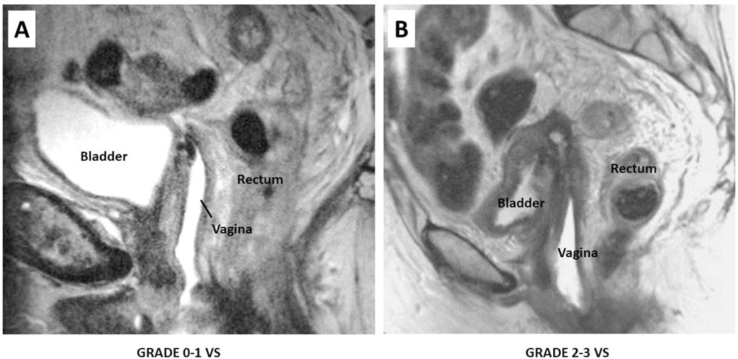
Purpose: Guidelines for the care of women undergoing pelvic radiation therapy (RT) recommend vaginal dilator therapy (VDT) to prevent radiation-induced vaginal stenosis (VS); however, no standard protocol exists. This review seeks to update our current state of knowledge concerning VS and VDT in radiation oncology.
Methods and materials: A comprehensive literature review (1972-2017) was conducted using search terms "vaginal stenosis," "radiation," and "vaginal dilator." Information was organized by key concepts including VS definition, time course, pathophysiology, risk factors, and interventions.
Results: VS is a well-described consequence of pelvic RT, with early manifestations and late changes evolving over several years. Strong risk factors for VS include RT dose and volume of vagina irradiated. Resultant vaginal changes can interfere with sexual function and correlational studies support the use of preventive VDT. The complexity of factors that drive noncompliance with VDT is well recognized. There are no prospective data to guide optimal duration of VDT, and the consistency with which radiation oncologists monitor VS and manage its consequences is unknown.
Conclusions: This review provides information concerning VS definition, pathophysiology, and risk factors and identifies domains of VDT practice that are understudied. Prospective efforts to monitor and measure outcomes of patients who are prescribed VDT are needed to guide practice.
Copyright © 2019 American Society for Radiation Oncology. All rights reserved.
Conflict of interest statement
Figures


References
-
- Hartman P, Diddle AW. Vaginal stenosis following irradiation therapy for carcinoma of the cervix uteri. Cancer 1972;30:426–429. - PubMed
-
- International guidelines on vaginal dilation after pelvic radiotherapy. Produced by the International Clinical Guideline Group, chaired by Dr Tracie Miles, President, National Forum of Gynaecological Oncology Nurses, UK. https://owenmumford.com/us/wpcontent/uploads/sites/3/2014/11/Dilator-Bes...
-
- Khor TH, Tuan JK, Hee SW, et al. Radical radiotherapy with high-dose-rate brachytherapy for uterine cervix cancer long-term results. Australas Radiol 2007;51:570–577. - PubMed
-
- Fraunholz IB, Schopohl B, Böttcher HD Management of radiation injuries of vulva and vagina. Strahlenther Onkol 1998;174 Suppl 3:90–2. - PubMed
-
- Nori D, Merimsky O, Batata M, et al. Postoperative high dose-rate intravaginal brachytherapy combined with external irradiation for early stage endometrial cancer: a long-term follow-up. Int J Radiat Oncol Biol Phys 1994;30:831–837. - PubMed
Publication types
MeSH terms
Grants and funding
LinkOut - more resources
Full Text Sources
Medical
