

Antibiotic modulation of mucins in otitis media; should this change our approach to watchful waiting?
- PMID: 31302575
- PMCID: PMC6742428
- DOI: 10.1016/j.ijporl.2019.07.002
Antibiotic modulation of mucins in otitis media; should this change our approach to watchful waiting?
Abstract
Background: Gel-forming mucins (GFMs) play important roles in otitis media (OM) pathogenesis. Increased mucin expression is activated by pathogens and proinflammatory cytokines. Bacterial biofilms influence inflammation and resolution of OM and may contribute to prolonged mucin production. The influence of specific pathogens on mucin expression and development of chronic OM with effusion (OME) remains an area of significant knowledge deficit.
Objectives: To assess the relationship between GFM expression, specific pathogens, middle ear mucosal (MEM) changes, biofilm formation, and antibiotic utilization.
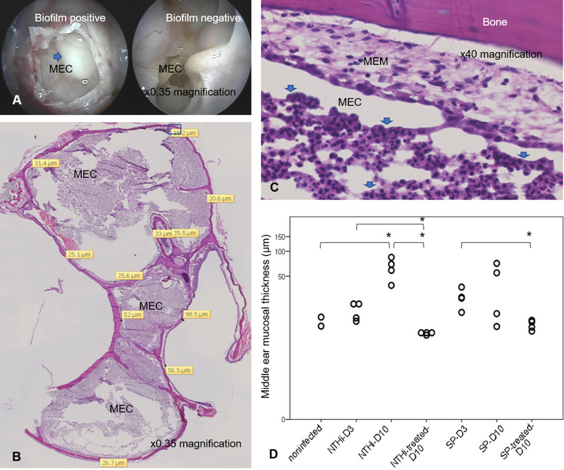
Methods: Mixed gender chinchillas were inoculated with nontypeable Haemophilus influenzae (NTHi) strain 86028NP or Streptococcus pneumoniae (SP) strain TIGR4 via transbulla injection. Antibiotic was administered on day 3-5 post inoculation. GFM expression was measured by quantitative PCR. Biofilm formation was identified and middle ear histologic changes were measured.
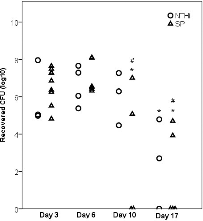
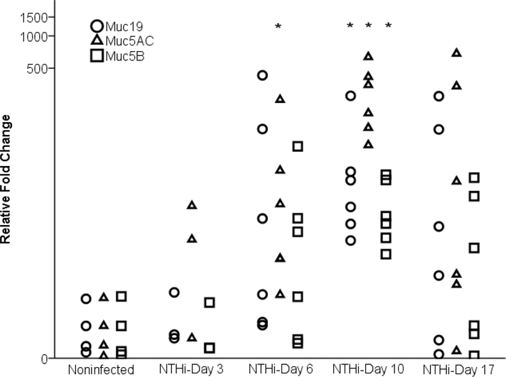
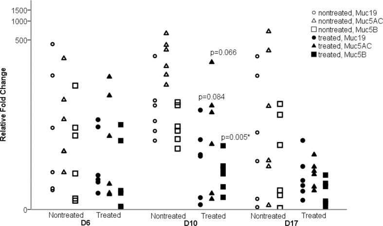
Results: SP infection resulted in higher incidence of biofilm and ME effusion compared with NTHi infection. However, NTHi persisted in the ME longer than SP with no substantive bacterial clearance detected on day 10 compared with complete bacterial clearance on day 10 for 50-60% of the SP-infected chinchillas. Both infections increased MEM inflammatory cell infiltration and thickening. NTHi upregulated the Muc5AC, Muc5B and Muc19 expression on day 10 (p = 0.0004, 0.003, and 0.002 respectively). SP-induced GFM upregulations were trended toward significant. In both NTHi and SP infections, the degree of GFM upregulation had a direct relationship to increased MEM hypertrophy, inflammatory cell infiltration and biofilm formation. Antibiotic treatment reduced the incidence of ME effusion and biofilm, limited the MEM changes and reversed the GFM upregulation. In NTHi infection, the rate of returning to baseline level of GFMs in treated chinchillas was quicker than those without treatment.
Conclusions: In an animal model of OM, GFM genes are upregulated in conjunction with MEM hypertrophy and biofilm formation. This upregulation is less robust and more quickly ameliorated to a significant degree in the NTHi infection with appropriate antibiotic therapy. These findings contribute to the understanding of pathogen specific influences on mucin expression during OM pathogenesis and provide new data which may have implications in clinical approach for OM treatment.
Keywords: Antibiotic treatment; Gel-forming mucins; Otitis media.
Copyright © 2019 Elsevier B.V. All rights reserved.
Conflict of interest statement
None of the authors of this manuscript have any financial or non-financial competing interests to disclose.
Figures
References
-
- Bluestone CD. Modern management of otitis media. Pediatr Clin North Am. 36(6) (1989):137–187. - PubMed
-
- Klein JO. Otitis media. Clin Infect Dis. 19(5) (1994):823–833. - PubMed
-
- Williamson IG, Dunleavey J, Bain J, Robinson D. The natural history of otitis media with effusion—a three-year study of incidence and prevalence of abnormal tympanograms in four South West Hampshire infant and first schools. J Laryngol Otol. 108(11) (1994):930–934. - PubMed
-
- Lin J, Tsuprun V, Kawano H, et al. Characterization of mucins in human middle ear and Eustachian tube. Am J Physiol Lung Cell Mol Physiol. 280(6) (2001):L1157–1167. - PubMed
-
- Kerschner JE. Mucin gene expression in human middle ear epithelium. Laryngoscope. 117(9) (2007):1666–1676. - PubMed
MeSH terms
Substances
Grants and funding
LinkOut - more resources
Full Text Sources
Medical
