. 2019 Nov;78(11):1524-1535.
doi: 10.1136/annrheumdis-2019-215543. Epub 2019 Jul 13.

Fexofenadine inhibits TNF signaling through targeting to cytosolic phospholipase A2 and is therapeutic against inflammatory arthritis

Affiliations

Fexofenadine inhibits TNF signaling through targeting to cytosolic phospholipase A2 and is therapeutic against inflammatory arthritis

Ronghan Liu et al. Ann Rheum Dis. 2019 Nov.

Abstract

Objective: Tumour necrosis factor alpha (TNF-α) signalling plays a central role in the pathogenesis of various autoimmune diseases, particularly inflammatory arthritis. This study aimed to repurpose clinically approved drugs as potential inhibitors of TNF-α signalling in treatment of inflammatory arthritis.

Methods: In vitro and in vivo screening of an Food and Drug Administration (FDA)-approved drug library; in vitro and in vivo assays for examining the blockade of TNF actions by fexofenadine: assays for defining the anti-inflammatory activity of fexofenadine using TNF-α transgenic (TNF-tg) mice and collagen-induced arthritis in DBA/1 mice. Identification and characterisation of the binding of fexofenadine to cytosolic phospholipase A2 (cPLA2) using drug affinity responsive target stability assay, proteomics, cellular thermal shift assay, information field dynamics and molecular dynamics; various assays for examining fexofenadine inhibition of cPLA2 as well as the dependence of fexofenadine's anti-TNF activity on cPLA2.

Results: Serial screenings of a library composed of FDA-approved drugs led to the identification of fexofenadine as an inhibitor of TNF-α signalling. Fexofenadine potently inhibited TNF/nuclear factor kappa-light-chain-enhancer of activated B cells (NF-ĸB) signalling in vitro and in vivo, and ameliorated disease symptoms in inflammatory arthritis models. cPLA2 was isolated as a novel target of fexofenadine. Fexofenadine blocked TNF-stimulated cPLA2 activity and arachidonic acid production through binding to catalytic domain 2 of cPLA2 and inhibition of its phosphorylation on Ser-505. Further, deletion of cPLA2 abolished fexofenadine's anti-TNF activity.

Conclusion: Collectively, these findings not only provide new insights into the understanding of fexofenadine action and underlying mechanisms but also provide new therapeutic interventions for various TNF-α and cPLA2-associated pathologies and conditions, particularly inflammatory rheumatic diseases.

Keywords: TNF-α; cPLA2; fexofenadine; inflammation; inflammatory arthritis.


Conflict of interest statement

Competing interests: None declared.

Figures
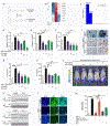
Fig. 1.
Fig. 1.. Fexofenadine acts as the antagonists of TNF-α and inhibit TNF-α signaling and activity.
a. The molecular structure of Fexofenadine (FFD) and Terfenadine (TFD). CYP3A4, the major enzyme responsible for the metabolic process, is indicated. b. BMDMs were treated without or with (10ng/ml) in absence or presence of FFD (10 μM) for 24 hours. Total RNA was extracted for RNA-seq. A few typical TNF-α inducible genes that were suppressed by FFD were presented. c. Transcription factor enrichment analysis from RNA-seq results, indicating the decreased gene expressions resulted from the suppressed activity of transcription factors NF-ĸB1 and RELA by FFD. d–f. BMDMs were treated with or without (10 ng/ml) in absence or presence of FFD (1 μM, 10 μM)/TFD (0.1 μM, 1 μM) for 24 hours. mRNA expressions of IL-1β, IL-6 and Nos-2 were tested by qRT-PCR. g–h. BMDMs were treated without or with TNF-α (10 ng/ml) in absence or presence of FFD (1μM, 10μM)/TFD (0.1μM, 1μM) for 48 hours. The levels of IL-1β and IL-6 in supernatant were detected by ELISA. i. BMDMs were treated with M-CSF (10 ng/ml) for 3 days, then cultured with RANKL (100ng/ml) and TNF-α (10 ng/ml) with or without FFD (10 μM) or TFD (1 μM) for 4 days and TRAP staining was performed. Scale bar, 100μm. j. TNF-tg/NF-kB-Luc mice were applied to examine the anti-TNF effects of FFD/TFD in vivo. After FFD (2 or 10 mg/kg) and TFD (10 or 50 mg/kg) were orally administrated for 7 days, luciferase signals were detected by IVIS system. k. BMDMs were with treated with TNF-α (10 ng/ml) in the absence or presence of FFD (10 μM)/or TFD (1 μM) for various time points, as indicated. Cytoplasmic (CE) and nuclear extractions (NE) were examined by Western blot with anti-p65 antibody. l. BMDMs were cultured with TNF-α (10 ng/ml) in the absence or presence of FFD (10 μM) or TFD (1 μM) for 6 hours. Immunofluorescence cell staining was performed to visualize the subcellular localization of p65. DAPI was used to stain the nucleus. Scale bar, 25μm. m. p65 DNA binding activity was tested by ELISA. Excess amounts (100X) of WT and mutant Oligo were used as positive and negative control respectively. (* p<0.05, ** p<0.01, ***p<0.001).
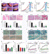
Fig. 2.
Fig. 2.. Fexofenadine prevents the spontaneous development of inflammatory arthritis in TNF transgenic mice.
a–h. TNF-tg mice (n=6) were orally administered Fexofenadine (FFD, 10 mg/kg), Terfenadine(TFD, 50 mg/kg), or Methotrexate (MTX, 2 mg/kg, serving as a positive control) daily beginning at 8-weeks of age and continuing for a total of 13 weeks. During this period, treatment was halted at 17-week point indicated by red arrow and resumed at 19-week point indicated by green arrow. a. Representative images of front paws and hind paws. b–c. Swelling score. d. H&E staining and quantification of histological score of knee and ankle samples. e. TRAP staining of paw and skull samples. f. Safranin O staining of knee and ankle samples. g–h. Serum levels of IL-1β and IL-6, assayed by ELISA. i–j. Therapeutic effects of FFD/TFD were tested by treating the TNF-tg mice with average swelling score reached around 8 points (n=6). Swelling scores were recorded weekly. (* p<0.05, ** p<0.01, ***p<0.001). (Scale bar, 100μm)
Fig. 3.
Fig. 3.. Fexofenadine prevents the onset and progression of collagen-induced arthritis.
a–j. Collagen-induced arthritis (CIA) model of DBA/1J mice was used to test prevention effects of Fexofenadine (FFD) and Terfenadine (TFD), (n=8). FFD (10 mg/kg), TFD (50 mg/kg) and MTX (2 mg/kg) were orally delivered daily beginning 18 days after immunization. a. The representative images of front paws and hind paws. b. Paw thickness. c. Clinical score of CIA. d. The incidence rate of arthritis. e. H&E staining and quantification of histological score of ankle samples. f. TRAP staining of ankle samples. g. microCT of ankles. h. Safranin O staining of ankle samples. i–j. The serum levels of IL-1β and IL-6 in CIA models. k–p. To examine the dosage-dependent therapeutic effects of FFD/TFD, CIA mice were treated with various dose of FFD or TFD, as indicated. FFD, TFD, MTX and vehicle were delivered after the clinical score reached approximately 5 points. k. The clinical score of FFD treated mice. l. The clinical score of TFD treated mice. m–p. The serum levels of IL-1β and IL-6. (n=8) (* p<0.05, ** p<0.01, ***p<0.001). (Scale bar, 100μm)
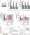
Fig. 4.
Fig. 4.. Fexofenadine’s anti-TNF activity is H1R1-independant.
a–b. The anti-TNF activity of Terfenadine (TFD) and Fexofenadine (FFD) does not depend on H1R1. a. Immunoblotting analysis to examine the knockdown efficacy of siRNA against H1R1. b. RAW264.7 cells transfected with scrambled control siRNA (scRNAi) or H1R1 RNAis were treated with or without TNFα (10ng/ml) in absence or presence of FFD (10μM)/TFD(1μM) for 48 hours. The levels of IL-1β and IL-6 in the medium were detected by ELISA. c–d. Comparison of the anti-TNF activity between Terfenadine (TFD)/Fexofenadine (FFD) and other known H1R1 inhibitors. BMDM cells were treated without or with TNF-α(10ng/ml) in absence or presence of various H1R1 inhibitor, as indicated, for 48 hours. The levels of IL-1β and IL-6 in medium were detected by ELISA. e–g. Terfenadine (TFD) and Fexofenadine (FFD) do not affect the binding of TNF-α and TNFR1 and to the cell surface. e. Solid phase binding was used to reveal the dose-dependent binding of TNF-α to TNFR1. f. The binding of TNF-α to TNFR1 in the presence of DMSO (negative control), FFD or TFD was also analyzed by solid phase binding. g. RAW264.7 cells were incubated with biotin-labelled TNF-α in the absence or presence of TNF antibody (positive control), FFD (10 μM) or TFD (1 μM) for overnight, then cells were analyzed by flow cytometry. (* p<0.05, ** p<0.01, ***p<0.001).
Fig. 5.
Fig. 5.. cPLA2 is a novel target of Fexofenadine.
a. Silver staining of DARTS assay. b. Coomassie blue staining of DARTS assay. The band with molecular weight around 80 kDa protected by Fexofenadine (FFD)/Terfenadine (TFD) was indicated by arrow. c. Adapted image of a mass spectra for PLA2G4A, encoding cPLA2. d. DARTS and Western blot to confirm FFD/TFD’s binding targets. e. CETSA melt response and associated curve. f. Isothermal dose response (ITDR) and its curve. g. IFD simulated binding complexes of cPLA2-FFD and cPLA2-TFD, respectively. cPLA2 is shown by surface representation (grey). FFD and TFD are shown by CPK representation with the atoms colored as carbon–violet (FFD) or cyan (TFD), oxygen–red, nitrogen–blue, hydrogen–white (only polar hydrogens of ligand are shown). h–i. Docked poses of FFD and TFD in cPLA2, respectively, predicted by IFD. FFD and TFD are shown as ball and stick model with the same atom color scheme. Important amino acids are depicted as sticks with the same color scheme except that carbon atoms are represented in grey. Only polar hydrogens are shown. Dotted yellow lines indicate hydrogen-bonding interactions. Values of the relevant distances are given in Å. j. DARTS assay with serial deletion constructs encoding Flag-tagged mutants of cPLA2. cPAL2 (aa 1–750), cPLA2 (aa 126–750), cPLA2 (aa 406–750), cPLA2 (aa 1–479), cPLA2 (aa 1–144). 293T cells were transfected with Flag-tagged mutants of cPLA2 plasmids, as indicated. DARTS assay samples were detected by Flag antibody. k. DARTS assay for Ser-505 point mutant of cPLA2. The Ser-505 of cPLA2 was substituted with Ala505. 293T cells were transfected with the point mutant plasmid and DARTS was performed. Point mutated cPLA2 was detected by Flag antibody.
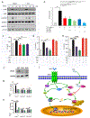
Fig. 6.
Fig. 6.. Fexofenadine inhibits TNF activity through binding to the catalytic domain 2 of cPLA2 and inhibition of the phosphorylation of cPLA2 on Ser-505.
a. Fexofenadine (FFD) inhibits the phosphorylation of cPLA2 on Ser-505. BMDM cells were treated with TNF-α (10 ng/ml) in the absence or presence of FFD (10 μM) for various time points, as indicated. p-p38, t-p38, p-ERK1/2, t-ERK1/2, p-cPLA2 (specifically for phosphorylated Ser-505), t-cPLA2 were detected by Western blot with corresponding antibodies. b. Fexofenadine inhibits TNF-induced cPLA2 activity in living cells. RAW264.7 cells transfected with an expression plasmid encoding cPLA2 were treated with TNF-α and ATK, or Terfenadine (TFD), or FFD overnight. Cells lysate was used for cPLA2 activity analysis. c. FFD inhibits TNF-induced arachidonic acid (AA) production. The AA levels in BMDMs without or with TNF-α (10 ng/ml) in absence or presence of FFD or TFD for 48 hours were examined using a commercial ELISA kit. ATK was used as a positive control. d–e. Addition of AA abolished FFD inhibition of TNF-induced cytokine release. BMDMs were treated with TNF-α (10 ng/ml), AA (10 μM), and FFD (10 μM)/TFD (1 μM), as indicated. The levels of IL-1β and IL-6 were detected by ELISA. f. Knock out efficiency of cPLA2 using CRISPR-Cas9 technique in RAW264.7 cells, assayed by Western blot. Two individual knockout clones (KO1 and KO2) were employed. g–h. Deletion of cPLA2 abolished FFD inhibition of TNF-induced cytokine release. WT and cPLA2 KO RAW264.7 cells were treated without or with TNF-α (10 ng/ml) in absence or presence of FFD (10 μM)/TFD (1 μM) for 48 hours. The levels of IL-1β and IL-6 in medium were detected by ELISA. (* p<0.05, ** p<0.01, ***p<0.001). i. A proposed model for explaining the anti-TNF activity of FFD through targeting cPLA2 pathway.

Similar articles

Cited by

References

    1. McKinney EF, Lee JC, Jayne DR, et al. T-cell exhaustion, co-stimulation and clinical outcome in autoimmunity and infection. Nature 2015, 523(7562):612–616. - PMC - PubMed
    1. Barturen G, Beretta L, Cervera R, et al. Moving towards a molecular taxonomy of autoimmune rheumatic diseases. Nature reviews Rheumatology 2018, 14(2):75–93. - PubMed
    1. Surolia I, Pirnie SP, Chellappa V, et al. Functionally defective germline variants of sialic acid acetylesterase in autoimmunity. Nature 2010, 466(7303):243–247. - PMC - PubMed
    1. Brenner D, Blaser H, Mak TW. Regulation of tumour necrosis factor signalling: live or let die. Nat Rev Immunol 2015, 15(6):362–374. - PubMed
    1. Croft M, Benedict CA, Ware CF. Clinical targeting of the TNF and TNFR superfamilies. Nat Rev Drug Discov 2013, 12(2):147–168. - PMC - PubMed

Publication types

MeSH terms