

Increased extracellular volume in the liver of pediatric Fontan patients
- PMID: 31303178
- PMCID: PMC6628496
- DOI: 10.1186/s12968-019-0545-4
Increased extracellular volume in the liver of pediatric Fontan patients
Abstract
Background: Patients with single ventricle physiology are at increased risk for developing liver fibrosis. Its extent and prevalence in children with bidirectional cavopulmonary connection (BCPC) and Fontan circulation are unclear. Extracellular volume fraction (ECV), derived from cardiovascular magnetic resonance (CMR) and T1 relaxometry, reflect fibrotic remodeling and/or congestion in the liver. The aim of this study was to investigate whether pediatric patients with single ventricle physiology experience increased native T1 and ECV as markers of liver fibrosis/congestion.
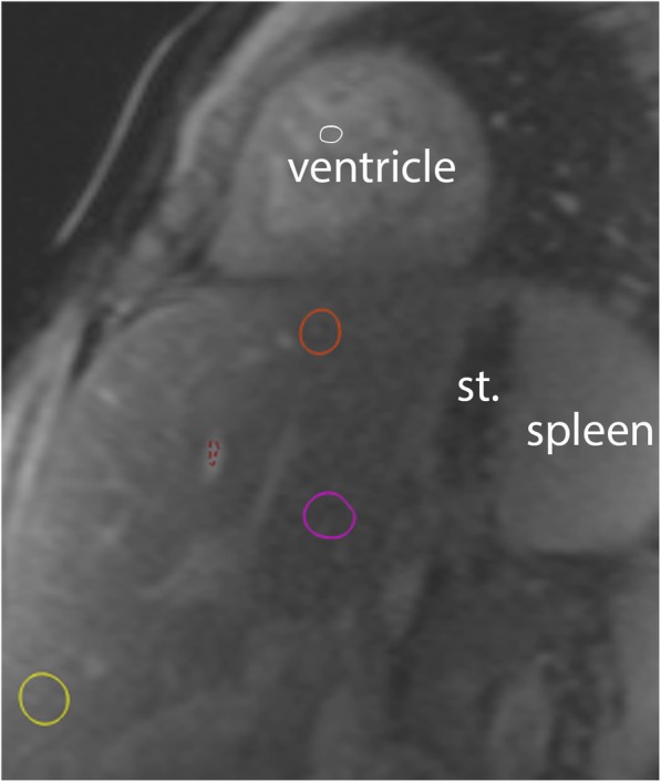
Methods: Hepatic native T1 times and ECV, using a cardiac short axis modified Look-Locker inversion recovery sequence displaying the liver, were measured retrospectively in children with BCPC- and Fontan circulations and compared to pediatric controls.
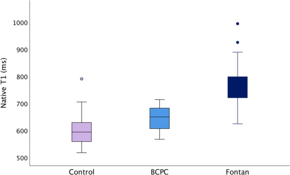
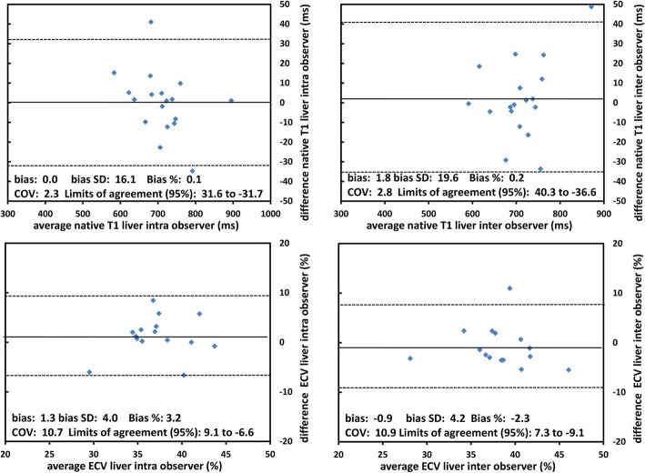
Results: Hepatic native T1 time were increased in Fontan patients (n = 62, 11.4 ± 4.4 years, T1 762 ± 64 ms) versus BCPC patients (n = 20, 2.8 ± 0.9 years, T1 645 ± 43 ms, p = 0.04). Both cohorts had higher T1 than controls (n = 44, 13.7 ± 2.9 years, T1 604 ± 54 ms, p < 0.001 for both). ECV was 41.4 ± 4.8% in Fontan and 36.4 ± 4.8% in BCPC patients, respectively (p = 0.02). In Fontan patients, T1 values correlated with exposure to cardiopulmonary bypass time (R = 0.3, p = 0.02), systolic and end diastolic volumes (R = 0.3, p = 0.04 for both) and inversely with oxygen saturations and body surface area (R = -0.3, p = 0.04 for both). There were no demonstrable associations of T1 or ECV with central venous pressure or age after Fontan.
Conclusion: Fontan and BCPC patients have elevated CMR markers suggestive of hepatic fibrosis and/or congestion, even at a young age. The tissue changes do not appear to be related to central venous pressures.
Trial registration: Retrospectively registered data.
Keywords: Cardiovascular magnetic resonance; Fontan circulation; Liver cirrhosis; Single ventricle; T1 mapping.
Conflict of interest statement
The authors declare that they have no competing interests.
Figures
Similar articles
-
Hepatic magnetic resonance T1-mapping and extracellular volume fraction compared to shear-wave elastography in pediatric Fontan-associated liver disease.Pediatr Radiol. 2021 Jan;51(1):66-76. doi: 10.1007/s00247-020-04805-y. Epub 2020 Oct 9. Pediatr Radiol. 2021. PMID: 33033916 Free PMC article.
-
Cardiac and Liver Fibrosis Assessed by Multiparametric MRI in Patients with Fontan Circulation.Pediatr Cardiol. 2025 Apr;46(4):966-975. doi: 10.1007/s00246-024-03522-9. Epub 2024 May 21. Pediatr Cardiol. 2025. PMID: 38771376 Free PMC article.
-
Myocardial fibrosis, diastolic dysfunction and elevated liver stiffness in the Fontan circulation.Open Heart. 2020 Oct;7(2):e001434. doi: 10.1136/openhrt-2020-001434. Open Heart. 2020. PMID: 33109703 Free PMC article.
-
Central Venous Waveform Patterns in the Fontan Circulation Independently Contribute to the Prediction of Composite Survival.Pediatr Cardiol. 2024 Dec;45(8):1617-1626. doi: 10.1007/s00246-023-03268-w. Epub 2023 Sep 23. Pediatr Cardiol. 2024. PMID: 37773462 Free PMC article. Review.
-
Surveillance for liver complications after the Fontan procedure.Congenit Heart Dis. 2017 Mar;12(2):124-132. doi: 10.1111/chd.12446. Epub 2017 Jan 31. Congenit Heart Dis. 2017. PMID: 28140526 Review.
Cited by
-
Efficacy Analysis of Double-Low Dynamic Contrast-Enhanced CT and Hepatic Extracellular Volume Fraction in the Diagnosis of Liver Fibrosis.Contrast Media Mol Imaging. 2022 Aug 17;2022:8089914. doi: 10.1155/2022/8089914. eCollection 2022. Contrast Media Mol Imaging. 2022. PMID: 36072627 Free PMC article.
-
Hepatic magnetic resonance T1-mapping and extracellular volume fraction compared to shear-wave elastography in pediatric Fontan-associated liver disease.Pediatr Radiol. 2021 Jan;51(1):66-76. doi: 10.1007/s00247-020-04805-y. Epub 2020 Oct 9. Pediatr Radiol. 2021. PMID: 33033916 Free PMC article.
-
T1 mapping of the myocardium and liver in the single ventricle population.Pediatr Radiol. 2023 May;53(6):1092-1099. doi: 10.1007/s00247-022-05560-y. Epub 2022 Dec 21. Pediatr Radiol. 2023. PMID: 36539566
-
Cardiac and Liver Fibrosis Assessed by Multiparametric MRI in Patients with Fontan Circulation.Pediatr Cardiol. 2025 Apr;46(4):966-975. doi: 10.1007/s00246-024-03522-9. Epub 2024 May 21. Pediatr Cardiol. 2025. PMID: 38771376 Free PMC article.
-
Regional Elevation of Liver T1 in Fontan Patients.CJC Pediatr Congenit Heart Dis. 2023 Mar 15;2(3):134-142. doi: 10.1016/j.cjcpc.2023.03.004. eCollection 2023 Jun. CJC Pediatr Congenit Heart Dis. 2023. PMID: 37969352 Free PMC article.
References
MeSH terms
LinkOut - more resources
Full Text Sources
Medical
