. 2019 Aug 1;105(2):403-412.
doi: 10.1016/j.ajhg.2019.06.007. Epub 2019 Jul 11.

De Novo Variants Disturbing the Transactivation Capacity of POU3F3 Cause a Characteristic Neurodevelopmental Disorder

Affiliations

De Novo Variants Disturbing the Transactivation Capacity of POU3F3 Cause a Characteristic Neurodevelopmental Disorder

Lot Snijders Blok et al. Am J Hum Genet. .

Abstract

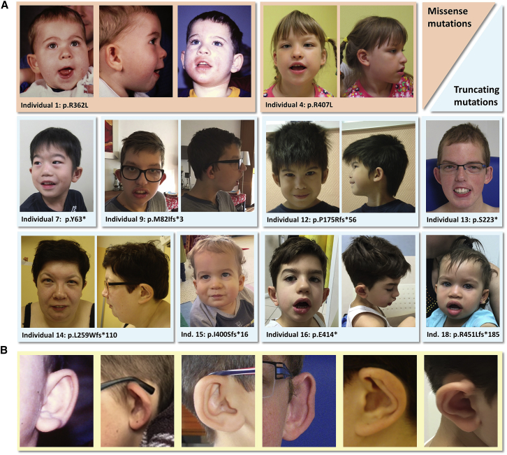
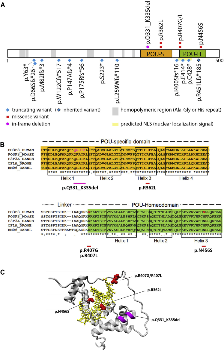
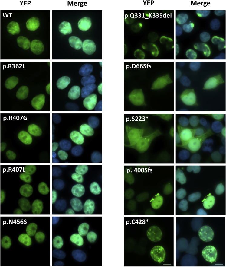
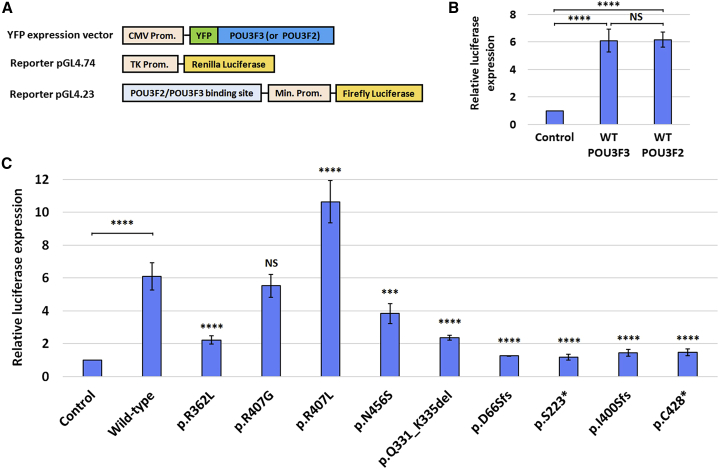
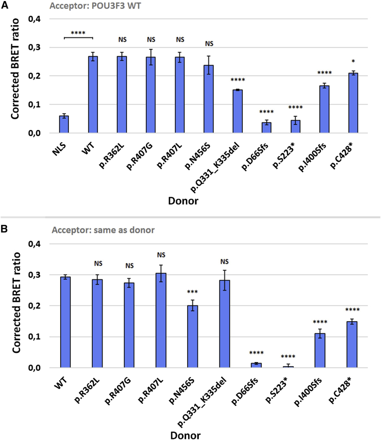
POU3F3, also referred to as Brain-1, is a well-known transcription factor involved in the development of the central nervous system, but it has not previously been associated with a neurodevelopmental disorder. Here, we report the identification of 19 individuals with heterozygous POU3F3 disruptions, most of which are de novo variants. All individuals had developmental delays and/or intellectual disability and impairments in speech and language skills. Thirteen individuals had characteristic low-set, prominent, and/or cupped ears. Brain abnormalities were observed in seven of eleven MRI reports. POU3F3 is an intronless gene, insensitive to nonsense-mediated decay, and 13 individuals carried protein-truncating variants. All truncating variants that we tested in cellular models led to aberrant subcellular localization of the encoded protein. Luciferase assays demonstrated negative effects of these alleles on transcriptional activation of a reporter with a FOXP2-derived binding motif. In addition to the loss-of-function variants, five individuals had missense variants that clustered at specific positions within the functional domains, and one small in-frame deletion was identified. Two missense variants showed reduced transactivation capacity in our assays, whereas one variant displayed gain-of-function effects, suggesting a distinct pathophysiological mechanism. In bioluminescence resonance energy transfer (BRET) interaction assays, all the truncated POU3F3 versions that we tested had significantly impaired dimerization capacities, whereas all missense variants showed unaffected dimerization with wild-type POU3F3. Taken together, our identification and functional cell-based analyses of pathogenic variants in POU3F3, coupled with a clinical characterization, implicate disruptions of this gene in a characteristic neurodevelopmental disorder.

Keywords: BRET assay; Brain-1; FOXP2; POU3F2; POU3F3; de novo variants; intellectual disability; luciferase reporter; speech/language disorder.


Conflict of interest statement

Z.P., S.T., and D.N.S. are full-time employees of Ambry Genetics. Exome sequencing is one of Ambry’s commercially available tests. The authors declare no other competing interests.

Figures

Figure 1
Figure 1
Facial Features of Ten Individuals with a Pathogenic POU3F3 Variant (A) All individuals in this picture have cupped and/or prominent and often low-set ears, except for individual 4. Other overlapping features are full lips, an open-mouth appearance, thick ear helices, a broad and bulbous nasal tip, hypertelorism, epicanthal folds, and peri-orbital fullness. (B) Magnification of the ear abnormalities in individuals 1, 9, 12, 13, 14, and 16, respectively.
Figure 2
Figure 2
Characterization of POU3F3 Variants (A) Linear representation of POU3F3 (UniProt: P20264) showing the location of variants from unrelated families in this cohort. There are twelve truncating variants (blue), five missense variants (red), and one in-frame deletion (magenta). POU-S (orange) is the POU-specific domain, and POU-H (green) is the POU-homeodomain. The shown NLS (nuclear localization signal) prediction is derived from cNLS Mapper. An overview with mutation details per subject is provided in Table S1. (B) Alignment of part of the POU3F3 amino acid sequence (using ClustalW) with orthologous sequences from the following species: Mus musculus, Danio rerio, Drosophila melanogaster, and Caenorhabditis elegans. Helix boundaries are defined as previously described. (C) Three-dimensional modeling of the functional domains of POU3F3 binding to a target DNA sequence (yellow). Amino acids that are affected by the missense variants are shown in red (wild-type side chains are depicted), and the location of the in-frame deletion is shown in magenta. A more detailed picture for two missense variants, p.Arg362Leu and p.Asn456Ser, can be found in Figure S2.
Figure 3
Figure 3
Subcellular Localization Direct fluorescence imaging of HEK293 cells expressing YFP-POU3F3 fusion proteins carrying different variants found in our cohort (green). The nuclei are stained with DAPI (blue). The scale bar = 10μm. Pictures showing the aberrant subcellular localization patterns in a larger amount of cells for the variants p.Gln331_Lys335del, p.S223, p.Ile400Serfs16, and p.Cys428 can be found in Figure S4.
Figure 4
Figure 4
Luciferase Assays (A) Expression constructs used in the luciferase assays: a YFP-fused POU3F3 or POU3F2 construct with a CMV promoter; a Firefly luciferase reporter construct with a minimal promoter and a preceding intronic FOXP2-derived binding site; and a control construct with Renilla luciferase under control of a TK promoter. (B) Results of luciferase assays with WT POU3F3 and WT POU3F2 and the reporter construct with the FOXP2-derived binding site. Values are expressed relative to the control and represent the mean ± SD of three independent experiments, each performed in triplicate (∗∗∗∗ = p < 0.0001 and NS = not significant, using one-way ANOVA and a post-hoc Tukey’s test). (C) Results of luciferase assay with WT POU3F3 and nine constructs with POU3F3 variants. Values are expressed relative to the control and represent the mean ± SD of three independent experiments, each performed in triplicate (∗∗∗ = p < 0.001; ∗∗∗∗ = p < 0.0001; and NS = not significant when compared to WT POU3F3 using one-way ANOVA and a post-hoc Dunnett’s test).
Figure 5
Figure 5
Bioluminescence Resonance Energy Transfer Assays (A) Bioluminescence resonance energy transfer (BRET) assays to measure interactions between WT POU3F3 and mutant POU3F3 constructs. Bars represent the corrected mean BRET ratio ± SD of three independent experiments performed in triplicate (∗∗∗∗ = p < 0.0001; = p < 0.05; and NS = not significant when compared to WT using one-way ANOVA and a post-hoc Tukey’s test). The NLS-donor construct is a Renilla luciferase construct with a nuclear localization signal. (B) BRET assays to measure homodimerization capacity of each mutant POU3F3 construct. Bars represent the corrected mean BRET ratio ± SD of three independent experiments performed in triplicate (∗∗∗∗ = p < 0.0001; ∗∗∗ = p < 0.001; and NS = not significant when compared to WT using one-way ANOVA and a post-hoc Tukey’s test)

References

    1. Sumiyama K., Washio-Watanabe K., Saitou N., Hayakawa T., Ueda S. Class III POU genes: Generation of homopolymeric amino acid repeats under GC pressure in mammals. J. Mol. Evol. 1996;43:170–178. - PubMed
    1. He X., Treacy M.N., Simmons D.M., Ingraham H.A., Swanson L.W., Rosenfeld M.G. Expression of a large family of POU-domain regulatory genes in mammalian brain development. Nature. 1989;340:35–41. - PubMed
    1. Hagino-Yamagishi K., Saijoh Y., Ikeda M., Ichikawa M., Minamikawa-Tachino R., Hamada H. Predominant expression of Brn-2 in the postmitotic neurons of the developing mouse neocortex. Brain Res. 1997;752:261–268. - PubMed
    1. McEvilly R.J., de Diaz M.O., Schonemann M.D., Hooshmand F., Rosenfeld M.G. Transcriptional regulation of cortical neuron migration by POU domain factors. Science. 2002;295:1528–1532. - PubMed
    1. Sugitani Y., Nakai S., Minowa O., Nishi M., Jishage K., Kawano H., Mori K., Ogawa M., Noda T. Brn-1 and Brn-2 share crucial roles in the production and positioning of mouse neocortical neurons. Genes Dev. 2002;16:1760–1765. - PMC - PubMed

Publication types