. 2019 Jul 22;29(14):2307-2321.e5.
doi: 10.1016/j.cub.2019.06.018. Epub 2019 Jul 11.

Loss of Function of Phosphodiesterase 11A4 Shows that Recent and Remote Long-Term Memories Can Be Uncoupled

Affiliations

Loss of Function of Phosphodiesterase 11A4 Shows that Recent and Remote Long-Term Memories Can Be Uncoupled

Katy Pilarzyk et al. Curr Biol. .

Abstract

Systems consolidation is a process by which memories initially require the hippocampus for recent long-term memory (LTM) but then become increasingly independent of the hippocampus and more dependent on the cortex for remote LTM. Here, we study the role of phosphodiesterase 11A4 (PDE11A4) in systems consolidation. PDE11A4, which degrades cyclic adenosine monophosphate (cAMP) and cyclic guanosine monophosphate (cGMP), is preferentially expressed in neurons of CA1, the subiculum, and the adjacently connected amygdalohippocampal region. In male and female mice, deletion of PDE11A enhances remote LTM for social odor recognition and social transmission of food preference (STFP) despite eliminating or silencing recent LTM for those same social events. Measurement of a surrogate marker of neuronal activation (i.e., Arc mRNA) suggests the recent LTM deficits observed in Pde11 knockout mice correspond with decreased activation of ventral CA1 relative to wild-type littermates. In contrast, the enhanced remote LTM observed in Pde11a knockout mice corresponds with increased activation and altered functional connectivity of anterior cingulate cortex, frontal association cortex, parasubiculum, and the superficial layer of medial entorhinal cortex. The apparent increased neural activation observed in prefrontal cortex of Pde11a knockout mice during remote LTM retrieval may be related to an upregulation of the N-methyl-D-aspartate receptor subunits NR1 and NR2A. Viral restoration of PDE11A4 to vCA1 alone is sufficient to rescue both the LTM phenotypes and upregulation of NR1 exhibited by Pde11a knockout mice. Together, our findings suggest remote LTM can be decoupled from recent LTM, which may have relevance for cognitive deficits associated with aging, temporal lobe epilepsy, or transient global amnesia.

Keywords: Kamin effect; cGMP; consolidation; cyclic nucleotide; memory lapse; phosphodiesterase; remote memory; system consolidation; systems consolidation; transient amnesia.


Conflict of interest statement

DECLARATION OF INTERESTS: The authors declare no competing interests

Figures

Figure 1.
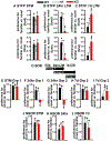
Figure 1.. Deletion of PDE11A produces transient amnesia for social transmission of food preference (STFP) and social odor recognition (SOR) but not non-social odor recognition (NSOR) in adult female and male mice.
A) Pde11a knockout (KO) mice (n=37) showed intact short-term memory (STM) for STFP (see Figure S1 for method detail), whereas, Pde11a heterozygous (HT) mice (n=33) showed a trend toward impaired STM relative to adult wild-type (WT) mice (n=32) (both failed equal variance; food: H(2)=5.51, P=0.063; time: H(2)=5.04, P=0.081). B) Pde11a heterozygous mice (n=24) showed strong recent long-term memory (LTM, 24 hours after training) relative to adult wild-type littermates (food, n=27; time, n=25); however, Pde11a knockout mice (food, n=30; time, 29) showed no recent LTM for STFP (food: F(2,75)=9.61, P=0.0002; time: F(2,72)=13.04, P=<0.0001). C) Despite these deficits at earlier time points, adult Pde11a heterozygous (n=24) and particularly adult Pde11a knockout mice (food, n=41; time, n=36) showed improved remote LTM for STFP (i.e., 7 days after training) relative to adult wild-type littermates (food, n=35; time, n=28) due to an apparent weakening of the memory in wild-type mice (failed equal variance; food: H(2)=13.48, P=0.0011; time: F(2,78)=8.84, P=0.0003). See Figure S2 for details regarding testing/retesting in STFP. D) STM, recent memory LTM, and remote LTM for SOR were assessed in 3 groups of mice. Group 1 was retested for 24 hour LTM on the same day Group 2 was retested for 7 day LTM. Mice in group 3 were trained/tested at a completely separate time. E) Group1 Pde11a knockout mice that showed normal STM (n=20/genotype; F(2,54)=0.61, P=0.55) went on to demonstrate F) impaired recent LTM (failed normality; H(2)=24.59, P<0.0001). G) Group 2 Pde11a knockout mice (n=21) tested for the first time 24 hours after training similarly showed impaired recent LTM relative to Pde11a heterozygous and wild-type littermates (n=20 each; failed normality; H(2)=31.17, P<0.0001), yet H) normal remote LTM (F(2,55)=0.43, P=0.65); see Figure S5 for replication). I) Surprisingly, Group 3 Pde11a heterozygous (n=13) and knockout mice (n=15) tested for the first time 7d after training actually showed slightly stronger remote LTM relative to wild-type mice (n=15; F(2,37)=3.60, P=0.037). In contrast, J) Pde11a deletion did not affect 2-bead NSOR memory (see Figure S5 for replication with 3-bead NSOR). Adult Pde11a wild-type, heterozygous and knockout mice showed equally strong STM (WT and KO, n=19; HT, n=11; F(1,43)= 0.258, P=0.774), K) recent LTM (WT, n=12; HT, n=10; KO, n=11; F(2,30)= 0.553, P=0.581) and L) remote LTM for NSOR (n=18/genotype, effect of bead: F(1,24)=22.55, P<0.001). Note, NSOR and STFP memories were only tested once. For NSOR, each cohort was naïve. See Table S1 for one-sample t-tests, Table S2 for other post hoc analyses, Figure S3 for novel vs. familiar plotted as a % of total (as previously reported [8]), and Figure S4 for total sniffing time that revealed a sex effect in SOR but no genotype effect. *significantly >0 (=memory), P<0.04-0.0001; Post hoc, #vs. WT, P≤0.035-0.0001. Data plotted as means ±SEMs.
Figure 2.
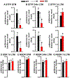
Figure 2.. Deletion of PDE11A produces transient amnesia for STFP and SOR but not NSOR in adolescent male and female mice.
A) Adolescent Pde11a heterozygous (n=15) and knockout mice (n=14) showed impaired STM for STFP (see Figure S1 for method detail) relative to adolescent wild-type littermates (n=16; food: F(2,39)=3.64, P=0.036; time: F(2,39)=4.13, P=0.023). B) This deficit recovers by 24 hours in adolescent Pde11a heterozygous mice (n=16) but not adolescent Pde11a knockout mice (n=24; WT, n=17; (both fail normality) food: H(2)=9.32, P=0.0094; time: H(2)=9.96, P=0.0069). Data represented in 2B top was previously reported in a different format in [8]. C) 7d after training, however, adolescent Pde11a knockout mice (n=26) showed intact remote LTM for STFP (WT, n=26; food: F(1,48)=2.17, P=0.148; time: F(1,48)=1.89, P=0.175). Although 7d STFP performance was slightly elevated in the adolescent knockout mice relative to wild-type littermates, the difference between genotypes was not statistically significant (e.g. food post hoc, P=0.095). The fact that adult Pde11a knockout mice showed significantly stronger 7d LTM for STFP relative to wild-type mice but adolescent mice did not may be due to the fact that performance of the adolescent wild-type mice did not weaken to the same extent as was seen in the adults (see Figure 1C). The weakening in adults is consistent with the fact that STFP memories are highly vulnerable to age-related decline in rodents [84]. D) Previously [8], we showed that adolescent Pde11a knockout mice also show no recent LTM for SOR; however, here we show adolescent Pde11a knockout mice (n=26) tested for the first time 7 days after training show remote LTM for SOR that is equivalent to that observed in heterozygous (n=32) and wild-type littermates (n=25; fails normality, H(2)=0.03, P=0.99). E) Separate cohorts of adolescent Pde11a wild-type, heterozygous and knockout mice also show equivalent NSOR when tested for short-term memory (n=18/genotype; F(1,32)=0.23, P=0.63), F) recent LTM 24 hr after training (WT, n=24; HT, n=17; KO, n=16; F(1,51)=2.01, P=0.15), and G) remote LTM 7 days after training (WT, n=17; HT, n=24; KO, n=18; F(1,56)=1.33, P=0.27). See Table S1 for one-sample t-tests, and Table S2 for other post hoc analyses. *significantly >0 (=memory), P<0.02-0.0001; Post hoc, #vs. WT, P=0.032-0.005. Data plotted as means ±SEMs.
Figure 3.
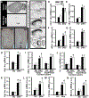
Figure 3.. Relative to adult wild-type littermates, Pde11a knockout mice show reduced activation of ventral CA1 (vCA1) during the retrieval of a recent LTM for STFP.
A) Pde11a4 mRNA and B) protein expression are restricted to CA1 stratum pyramidale (SP) and radiatum (SR), the subiculum (Sub), and the amygdalohippocampal area (just below CA1). Note the absence of PDE11A4 protein in CA2/CA3 as well as the lacunosum moleculare (LM). C) Autoradiographs for activity-regulated cytoskeleton associated protein (Arc) mRNA in home-caged controls (HC) and mice retrieving a 24hr STFP LTM (24hr, n =12/group; see Figure S6 for method detail). D) Pde11a knockout mice showed lower retrieval-induced Arc expression in vCA1 relative to wild-type littermates (F(3,41)=19.35, FDR-P<0.0001), but equivalent expression in E) ventral subiculum (vSub, F(3,44)=22.63, FDR-P<0.0001), F) dCA1 (F(3,44)=6.33, FDR-P=0.0002), G) dorsal subiculum (dSub, F(3,44)=46.05, FDR-P<0.0001), H) parasubiculum (PaS, F(3,44)=18.89, FDR-P<0.0001), I) the deep (H(3,44)=25.01, FDR-P=0.0002) and superficial layers (H(3,44)=28.38, FDR-P=0.0002) of medial entorhinal cortex (mEnt), J) the deep (H(3,44)=24.35, FDR-P=0.0004) and superficial layers (F(3,44)=33.20, FDR-P<0.0001) of visual cortex (V1), K,L) anterior cingulate subfields 1 and 2 (Cg1, H(3,42)=30.94, FDR-P<0.0001, Cg2: H(3,42)=20.23, FDR-P=0.0032), M) frontal association cortex (FrA, H(3,43)=28.64, FDR-P=0.0002), and N) motor cortex (M2, H(3,43)=22.20, FDR-P=0.003). See Table S2 for individual post hoc analyses. Post hoc, #vs. home cage within genotype (HC), P<0.01-0.001; Post hoc,*vs WT within behavioral group, P=0.002. Brightness and contrast of autoradiographic images adjusted for graphical clarity. Data plotted as means ±SEMs.
Figure 4.
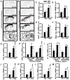
Figure 4.. Relative to adult wild-type littermates, Pde11a knockout mice show enhanced activation of extrahippocampal systems consolidation-related brain regions during retrieval of a remote LTM for STFP.
A,B) Arc mRNA autoradiographs (see Figure S6 for method detail) home-cage controls (HC) (n=7/genotype) and mice retrieving of a 7 day STFP LTM (7d, n =8/genotype). Relative to Pde11a wild-type mice, knockout mice showed equivalent retrieval-induced Arc expression in C) vCA1 (F(3,19)=7.82, FDR-P=0.001), D) vSub (F(3,19)=12.20, FDR-P=0.0001), E) dCA1 (F(3,19)=25.55, FDR-P<0.0001), and F) dSub (F(3,19)=29.84, FDR-P<0.0001). Relative to Pde11a wild-type mice, knockout mice showed significantly stronger retrieval-induced Arc expression in G) parasubiculum (PaS, F(3,29)=29.05, FDR-P<0.0001) and H) the superficial layer of medial entorhinal cortex (mEnt, F(3,29)=30.37, FDR-P<0.0001), but not the deep layers of Ent (F(3,19)=14.95, FDR-P<0.0001) nor I) visual cortex (V1, F(3,19)=17.48, FDR-P<0.0001). Relative to Pde11a wild-type mice, knockout mice also showed significantly stronger retrieval-induced Arc expression in J) anterior cingulate 1 (Cg1, F(3,29)=16.16, FDR-P<0.0001), K) Cg2 (F(3,29)=13.47, FDR-P<0.0001), and L) frontal association cortex (FrA, F(3,29)=10.15, FDR-P=0.0004), but not M) motor cortex (M2, F(3,19)=11.69, FDR-P=0.0002). See Table S2 for individual post hoc analyses. Post hoc, #vs HC within genotype, P<0.025-0.001; Post hoc, *vs WT within behavioral group, P=0.037-0.003. Brightness and contrast of autoradiographic images adjusted for graphical clarity. Data plotted as means ±SEMs.
Figure 5.
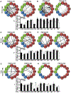
Figure 5.. Patterns of functional connectivity differ by both memory-retrieval period and Pde11a genotype in adult mice.
Correlational analyses were conducted comparing Arc expression between brain regions (see Figure S6 for method detail and Data S1 for each r and P-value). A,B,D,E,G,H) Black lines indicate significant correlations unique to that genotype, whereas, gray lines indicate correlations that are shared between genotypes for the same retrieval period (thick lines indicate P<0.05, thin lines indicate P=0.051-0.079—only shown when correlation reaches P<0.05 in other genotype). Solid lines indicate positive correlations, dashed lines indicate negative correlations. A,D,G) represent significant raw P-values as reported in [13] (with 66 inter-regional correlations per group and a significance cutoff of P<0.05, 3-4 false positives per group can be expected); whereas, B,E,H) represent only P-values that remain significant following FDR correction for multiple comparisons. C,F,I) represent a comparison of the strength of all inter-regional correlations for a given brain region and are graphed as mean ±SEM of the absolute value of the raw r (|r|). A-B) In the home cage, Pde11a knockout mice showed far less functional connectivity relative to wild-type mice, particularly in C) FrA (t(10)=3.15, raw-P=0.01 and FDR-P=0.04), Cg1 (t(10)=5.11, raw-P=0.0005 and FDR-P=0.0055), Cg2 (t(10)=2.37, raw-P=0.039; FDR-P=0.117) and M2 (t(10)=3.23, raw-P=0.009 and FDR-P=0.04). D-F) In striking contrast, there was not an overall reduction in functional connectivity in the PDE11A knockout versus wild-type mice when undergoing STFP retrieval 24 hours after training. Rather, there appears to be a shift in the functional connectivity of brain regions. For example, in wild-type mice, vCA1 correlates with sME; whereas, in knockout mice, vCA1 correlates with PaS. G-H) A similar shift in retrieval-induced functional connectivity is also seen between PDE11A knockout vs. wild-type mice 7 days after training, with some suggestion of a change in the functional connectivity of I) PaS (t(10)=2.72, raw-P=0.022 and FDR-P=0.26) and dSub (t(10)=2.36, raw-P=0.04 and FDR-P=0.37). vCA1—ventral CA1, vSub—ventral subiculum, dCA1—dorsal CA1, dSub—dorsal subiculum, PaS—parasubiculum, sME—superficial layer of medial entorhinal cortex, dME—deep layer of medial entorhinal cortex, V1—visual cortex, M2—motor cortex, FrA—frontal association cortex, Cg1—anterior cingulate cortex subfield 1, Cg2-- anterior cingulate cortex subfield 2. *both raw- and FDR-P≤0.04, ^only raw-P≤0.04.
Figure 6.
Figure 6.. Deletion of PDE11A increases expression of N-methyl-D-aspartate (NMDA) receptor subunits in prefrontal cortex but not ventral hippocampus of adult mice.
A) For purposes of this experiment, prefrontal cortex was defined as cortical tissue anterior to the caudate-putamen (brain shown from above; dissection indicated by perforated black line). B) Ventral hippocampus was defined as the lower 40-45% (hippocampus shown in the caudal plane and dissection indicated by the perforated black line). In prefrontal cortex, Pde11a knockout (KO) mice show C) increased expression of the NR1 (WT, n=36, KO, n=37, F(1,69)=4.67, P=0.034) and D) NR2A subunits (n=43/genotype, failed normality, Rank Sum Test, T(43,43)=1610, P=0.025) relative to wild-type littermates, but E) no change in expression of the NR2B subunit (WT, n=39, KO, n=40). In VHIPP, there was no difference between Pde11a knockout mice vs. wild-type littermates in terms of F) NR1 (n=17/genotype), G) NR2A (WT, n=30, KO, n=31), nor H) NR2B (WT, n=18, KO, n=19). *vs WT, P=0.034-0.025. See Figure S7 for images of full blots. Brightness and contrast of blot images adjusted for graphical clarity. Data plotted as means ±SEMs. Images in panels A and B from Allen Institute for Brain Sciences http://connectivity.brain-map.org./3d-viewer.
Figure 7.
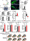
Figure 7.. Restoration of PDE11A4 in vCA1 of adult Pde11a knockout mice is sufficient to reverse social memory phenotypes without altering non-social memory.
A) Schematic of the custom lentiviral transfer vector used to drive expression of either eGFP-Pde11a4 or eGFP alone (green star = site of construct insertion). B) Expression in dorsal (DHIPP) and ventral hippocampus (VHIPP) of Pde11a knockout mice was confirmed by Western blot. C) Immunofluorescence 2 months following injection shows eGFP-PDE11A4 appropriately traffics throughout dendrites of stratum radiatum (SR) in CA1 (also see Figure S7). D) Relative to eGFP alone, eGFP-PDE11A4 expression significantly increases phosphorylation of the ribosomal protein S6 at residues 235/236 in VHIPP of Pde11a knockout mice (n=4/virus, t(6)=2.74, P=0.034), suggesting engagement of relevant signal transduction cascades [8]. E) Restoring eGFP-PDE11A4 expression also attenuates NR1 expression in PFC of Pde11a knockout mice (+eGFP, n=11, +eGFP-PDE11A4, n=10, F(1,17)=5.06, P=0.038) without changing F) NR2B expression, suggesting engagement of relevant circuits. G) Control knockout mice (n=20 GFP-treated + 1 sham) showed no recent LTM for STFP as expected (t(19)=1.55, P=0.14); however, PDE11A4-infected Pde11a knockout mice (vCA1+dCA1, n=16; vCA1 only, n=11) did show recent LTM (vCA1+dCA1: t(14)=5.54, P<0.0001; vCA1-only: t(10)=2.11, P=0.031). H) Also as expected, control Pde11a knockout mice (n=13) regained memory 7 days after training (t(12)=2.82, P=0.008); whereas, PDE11A4-infected Pde11a knockout mice (vCA1+dCA1, n=12, vCA1-only, n=6) acted like PDE11A wild-type mice showing no remote LTM for STFP (vCA1+dCA1: t(11)=0.67, P=0.26; vCA1-only: t(5)=0.53, P=0.69). I) Surprisingly, control knockout mice (n=20) showed a significant SOR LTM 24 hours after training (t(20)=4.86, P<0.0001), suggesting surgery partially rescued the SOR deficit normally observed in unsurgerized knockout mice (e.g., Figure 1G). That said, expression of eGFP-PDE11A4 in vCA1+dCA1 (n=20) or vCA1 alone (n=12) produced preference ratios that were twice as strong as that measured in the control group (failed normality; H(2)=16.92, P=0.0002) and nearly identical to those observed in wild-type mice (e.g., Figure 1G), suggesting a full rescue. It is not yet clear why surgerized knockouts here show a significant SOR memory 24 hours after training when unsurgerized knockouts do not. This effect may be related to severing of some vital circuit upon insertion of the needle or exposure to the anesthesia, but is not related to GFP expression as mock-surgerized knockout mice show a similar weak memory (see Figure S7I). To verify that viral expression of PDE11A4 affected only those behaviors impacted by genetic deletion, we lastly tested mice on NSOR. As expected, there was no effect of virus on recent LTM for NSOR using either the J) 3-bead protocol or K) 2-bead protocol. L) There was also no effect of the virus on remote LTM for NSOR. M) Schematic of working hypothesis. Deletion of PDE11A4 relocates memories to the cortex ahead of schedule at the expense of prematurely erasing/silencing the hippocampal trace, which temporarily “misplaces” the memory because retrieval vectors are not similarly updated in an expedited manner. The green arrow pointing from the hippocampus (in blue) to the prefrontal cortex along with the emergence of green coloration in prefrontal cortex indicates systems consolidation within the cortex, while the red line drawn from prefrontal cortex towards hippocampus and red coloration of the ventral hippocampus indicates silencing/erasure. Timing of retrieval vector updating is based on lesion studies summarized elsewhere [1]). See Table S1 for one-sample t-tests, and Table S2 for other post hoc analyses. LTR—long terminal repeat (*=mutated), HIV—human immunodeficiency virus, PGKp—phosphoglycerate kinase 1 promoter, eGFP—emerald green fluorescent protein, WPRE—woodchuck hepatitis virus post-transcriptional regulatory element, Amp-R—ampicillin resistance cassette, LM—lacunosum moleculare, SO—stratum oriens. Post hoc, #vs. GFP/control, P=0.034-0.0008; *significantly >0 (=memory), P≤0.03-0.0001. Data plotted as means ±SEMs.

Similar articles

Cited by

References

    1. Frankland PW, and Bontempi B (2005). The organization of recent and remote memories. Nat Rev Neurosci 6, 119–130. - PubMed
    1. Tonegawa S, Morrissey MD, and Kitamura T (2018). The role of engram cells in the systems consolidation of memory. Nat Rev Neurosci 19, 485–498. - PubMed
    1. Kitamura T, Ogawa SK, Roy DS, Okuyama T, Morrissey MD, Smith LM, Redondo RL, and Tonegawa S (2017). Engrams and circuits crucial for systems consolidation of a memory. Science 356, 73–78. - PMC - PubMed
    1. Tseng KY, Chambers RA, and Lipska BK (2009). The neonatal ventral hippocampal lesion as a heuristic neurodevelopmental model of schizophrenia. Behav Brain Res 204, 295–305. - PMC - PubMed
    1. Kogan JH, Frankland PW, and Silva AJ (2000). Long-term memory underlying hippocampus-dependent social recognition in mice. Hippocampus 10, 47–56. - PubMed

Publication types

Substances