

Radiomorphological Manifestations of Femoral and Tibial Cortical Bones at Different Stages of Limb Lengthening
- PMID: 31303674
- PMCID: PMC6590022
- DOI: 10.4103/ortho.IJOrtho_443_18
Radiomorphological Manifestations of Femoral and Tibial Cortical Bones at Different Stages of Limb Lengthening
Abstract
Background: There has been a lot of research done on Ilizarov's limb lengthening; however, very few publications focus on the quantitative assessment of the distractional bone regeneration in tibial and femur lengthening. Data regarding quality of the bone after lengthening are needed to consider the time of frame removal and develop a rehabilitation program.
Materials and methods: Computed tomography (CT) assessment of a parent bone was performed on 136 patients with limb length discrepancy and bone deformity of various etiologies before and after lengthening. Transosseous osteosynthesis technique with the Ilizarov's external fixation was used for limb lengthening and deformity correction in all the cases. A 64-slice scanner was used for CT assessments. Specific Roentgen-negative units of the Ilizarov apparatus and techniques for interpreting CT findings were employed for artifact-free densitometric assessment.
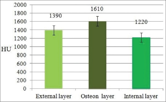
Results: Cortical density of the femur and tibia in patients with limb length discrepancy and bone deformity of various etiologies was shown to have differences as compared to the contralateral limb. The lengthening process was accompanied by decreased cortical density of the segment being lengthened, and the decrease in the density was greater in the areas adjacent to the distractional bone regeneration. The cortical structure underwent characteristic changes. Osteonal density of the cortical bone was higher in the norm and at long term followup as compared to the density of external and internal plates.
Conclusion: Cortical bone of the femur and tibia in patients with limb length discrepancy and bone deformity of various etiologies showed various preoperative local densities of external, internal, and osteon layers. The cortical bone demonstrated heterogenic structures with resorption areas of various magnitude and density, with minimal values at the boundary with regenerate bone during distraction and fixation with frame on and at short-term followup. Complete organotypical restructuring of the bone was shown to occur at a 1-to-3-year followup depending on the etiology of the disease and amount of lengthening performed.
Keywords: Corticotomy; Ilizarov; femur; limb lengthening; radiologic assessment; regenerate; tibia.
Conflict of interest statement
There are no conflicts of interest.
Figures


Similar articles
-
Reamed Intramedullary Nailing has an Adverse Effect on Bone Regeneration During the Distraction Phase in Tibial Lengthening.Clin Orthop Relat Res. 2016 Mar;474(3):816-24. doi: 10.1007/s11999-015-4613-2. Epub 2015 Oct 27. Clin Orthop Relat Res. 2016. PMID: 26507338 Free PMC article.
-
Limb lengthening and correction of deformity in the lower limbs of children with osteogenesis imperfecta.J Bone Joint Surg Br. 2004 Mar;86(2):259-65. doi: 10.1302/0301-620x.86b2.14393. J Bone Joint Surg Br. 2004. PMID: 15046443
-
Extensive limb lengthening in Ollier's disease: 25-year follow-up.Medicina (Kaunas). 2005;41(10):861-6. Medicina (Kaunas). 2005. PMID: 16272834
-
Retrograde magnetic internal lengthening nail for acute femoral deformity correction and limb lengthening.Expert Rev Med Devices. 2017 Oct;14(10):811-820. doi: 10.1080/17434440.2017.1378092. Epub 2017 Sep 17. Expert Rev Med Devices. 2017. PMID: 28893094 Review.
-
The use of flexible intramedullary nails in limb lengthening.Expert Rev Med Devices. 2017 Sep;14(9):741-753. doi: 10.1080/17434440.2017.1367284. Epub 2017 Aug 18. Expert Rev Med Devices. 2017. PMID: 28817981 Review.
References
-
- Diachkova GV, Neretin AS, Korabelnikov MA, Nizhechik SA. Radiological manifestations of bone tissue regeneration in treatment of patients with foot malformations (In Russian) Genii Ortopedii. 2005;4:98–101.
-
- Diachkova GV, Mikhailov ES, Erofeev SA, Diachkov SA, Diachkov KA. Radiological principles of assessment of distractional regenerate bone (In Russian) Travmatol Ortoped Protezirovanie Zapadnoy Sibiri. 2006;1:18–20.
-
- Diachkova GV, Mikhailov ES, Erofeev SA, Nizhechik SA, Korabelnikov MA. Qualitative and quantitative measurements of radiological distractional regenerate bone (In Russian) Genii Ortopedii. 2003;4:11–4.
-
- Shevtsov VI, Korabelnikov MA, Diachkova GV, Sukhodolova LV, Borzunov DY. CT quantitative assessment of reparative bone formation in experiment. Travmatol Ortoped Rossii. 2006;3:56–61.
LinkOut - more resources
Full Text Sources