Review
. 2019 Jun 28:15:1701-1716.
doi: 10.2147/NDT.S177804. eCollection 2019.

Stereoelectroencephalography in epilepsy, cognitive neurophysiology, and psychiatric disease: safety, efficacy, and place in therapy

Affiliations
Review

Stereoelectroencephalography in epilepsy, cognitive neurophysiology, and psychiatric disease: safety, efficacy, and place in therapy

Brett E Youngerman et al. Neuropsychiatr Dis Treat. .

Abstract

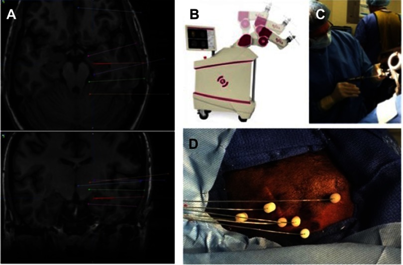
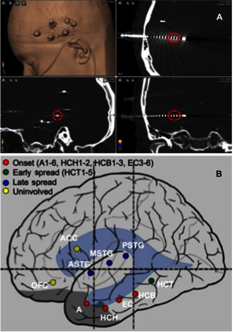
For patients with drug-resistant epilepsy, surgical intervention may be an effective treatment option if the epileptogenic zone (EZ) can be well localized. Subdural strip and grid electrode (SDE) implantations have long been used as the mainstay of intracranial seizure localization in the United States. Stereoelectroencephalography (SEEG) is an alternative approach in which depth electrodes are placed through percutaneous drill holes to stereotactically defined coordinates in the brain. Long used in certain centers in Europe, SEEG is gaining wider popularity in North America, bolstered by the advent of stereotactic robotic assistance and mounting evidence of safety, without the need for catheter-based angiography. Rates of clinically significant hemorrhage, infection, and other complications appear lower with SEEG than with SDE implants. SEEG also avoids unnecessary craniotomies when seizures are localized to unresectable eloquent cortex, found to be multifocal or nonfocal, or ultimately treated with stereotactic procedures such as laser interstitial thermal therapy (LITT), radiofrequency thermocoagulation (RF-TC), responsive neurostimulation (RNS), or deep brain stimulation (DBS). While SDE allows for excellent localization and functional mapping on the cortical surface, SEEG offers a less invasive option for sampling disparate brain areas, bilateral investigations, and deep or medial targets. SEEG has shown efficacy for seizure localization in the temporal lobe, the insula, lesional and nonlesional extra-temporal epilepsy, hypothalamic hamartomas, periventricular nodular heterotopias, and patients who have had prior craniotomies for resections or grids. SEEG offers a valuable opportunity for cognitive neurophysiology research and may have an important role in the study of dysfunctional networks in psychiatric disease and understanding the effects of neuromodulation.

Keywords: SEEG; cognitive neurophysiology; epilepsy surgery; psychiatric neurosurgery; stereoelectroencephalography.


Conflict of interest statement

The authors report no conflicts of interest in this work.

Figures

Figure 1
Figure 1
SEEG stereotactic planning and surgical technique. (A) Electrode trajectories are planned based on the pre-implantation hypothesis of the epileptogenic zone. We attempt to employ primarily orthogonal trajectories, avoid cortical vessels and sulci, and maximize gray matter sampling from gyral crowns, through sulcal cortex and depth of sulci, and to deep and medial targets. (B) After positioning and registration, the ROSA stereotactic robot is used to navigate to each trajectory in the operating room. (C) A small percutaneous incision and twist drill hole is made in line with the planned trajectory. (D) An anchor bolt is secured in the skull and the electrode passed to its premeasured depth and secured in the bolt. The process is repeated for each planned trajectory.
Figure 2
Figure 2
Localization of the epileptogenic zone. (A) A postoperative volumetric CT scan is obtained and used to localize each electrode contact in stereotactic space on the co-registered preoperative MRI. For each seizure recorded, the contacts involved are identified. (B) A summary of the contacts associated with seizure onset, early spread, and late spread is determined at an interdisciplinary epilepsy conference and used to plan further surgical intervention. Nomenclature is by convention. Each trajectory is named during the planning phase and numbers reflect the electrode contacts in order from deep to superficial (eg, A1–6=amygdala electrode contacts 1-6; HCH1–2=hippocampal head electrode contacts 1-2).

Similar articles

Cited by

References

    1. Brodie MJ, Barry SJ, Bamagous GA, Norrie JD, Kwan P. Patterns of treatment response in newly diagnosed epilepsy. Neurology. 2012;78(20):1548–1554. doi:10.1212/WNL.0b013e3182563b19 - DOI - PMC - PubMed
    1. Kwan P, Brodie MJ. Early identification of refractory epilepsy. N Engl J Med. 2000;342(5):314–319. doi:10.1056/NEJM200002033420503 - DOI - PubMed
    1. Kwan P, Arzimanoglou A, Berg AT, et al. Definition of drug resistant epilepsy: consensus proposal by the ad hoc task force of the ILAE commission on therapeutic strategies. Epilepsia. 2010;51(6):1069–1077. doi:10.1111/j.1528-1167.2009.02397.x - DOI - PubMed
    1. Englot DJ, Wang DD, Rolston JD, Shih TT, Chang EF. Rates and predictors of long-term seizure freedom after frontal lobe epilepsy surgery: a systematic review and meta-analysis. J Neurosurg. 2012;116(5):1042–1048. doi:10.3171/2012.1.JNS111620 - DOI - PubMed
    1. Piotrowska N, Winkler PA. Otfrid Foerster, the great neurologist and neurosurgeon from Breslau (Wroclaw): his influence on early neurosurgeons and legacy to present-day neurosurgery. J Neurosurg. 2007;107(2):451–456. doi:10.3171/JNS-07/08/0451 - DOI - PubMed