. 2018 Oct:2018:10.1109/ICSENS.2018.8589733.
doi: 10.1109/ICSENS.2018.8589733. Epub 2018 Dec 27.

Evaluation of High Frequency Piezoelectric Micromachined Ultrasound Transducers for Photoacoustic Imaging

Affiliations

Evaluation of High Frequency Piezoelectric Micromachined Ultrasound Transducers for Photoacoustic Imaging

Ajay Dangi et al. Proc IEEE Sens. 2018 Oct.

Abstract

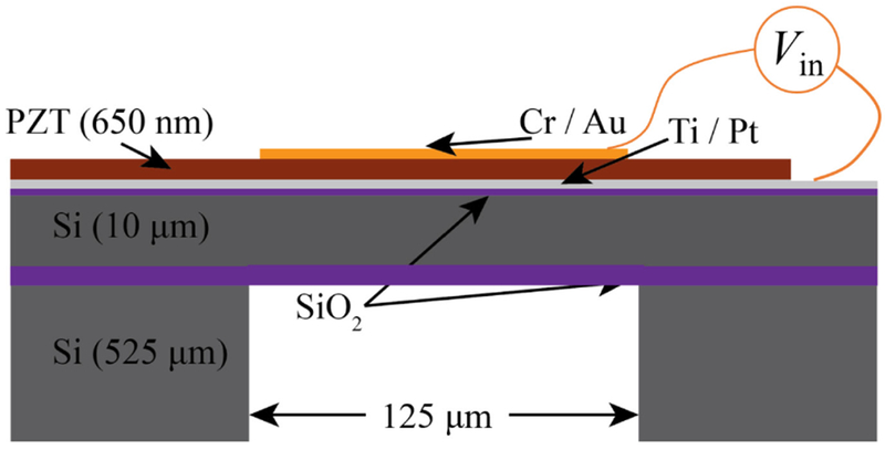
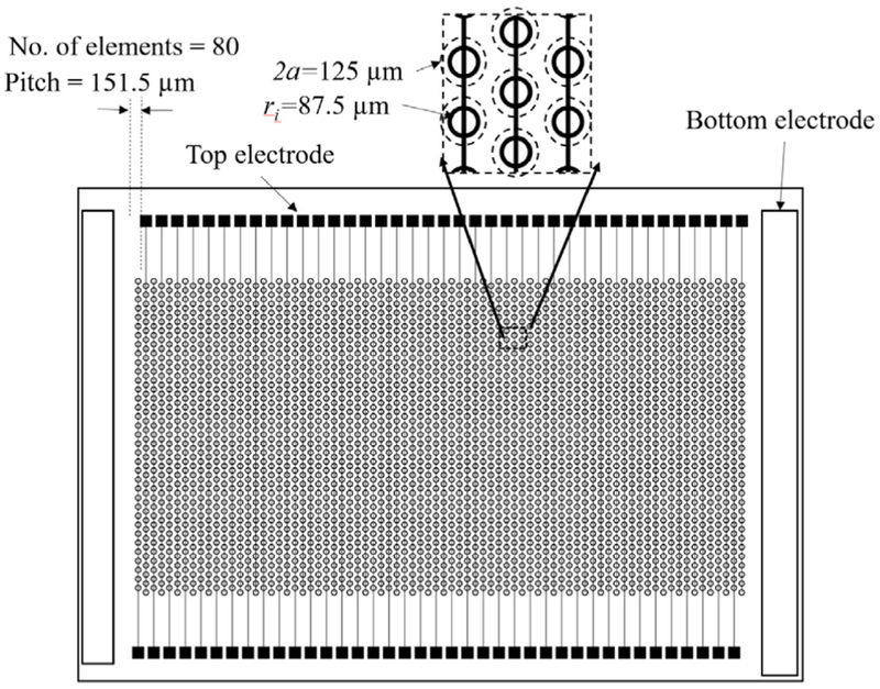
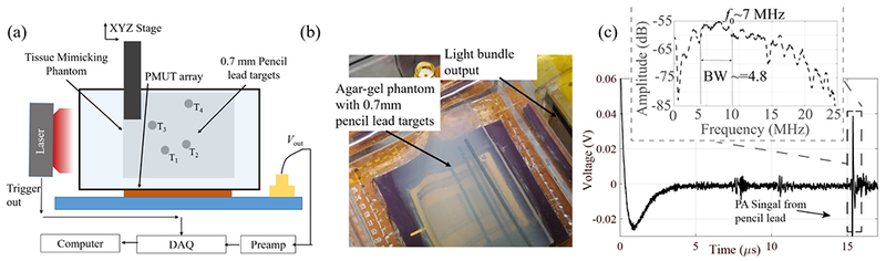
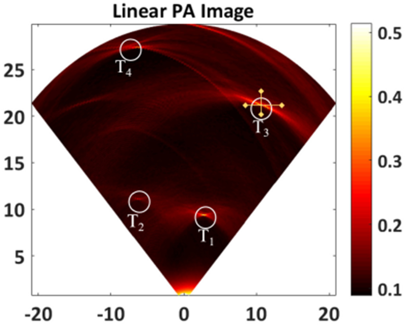
In this work, the design, fabrication, and characterization of piezoelectric micromachined ultrasound transducer (PMUT) arrays for photoacoustic imaging applications are reported. An 80-element linear PMUT array with each element having 53 PMUT cells of 125 μm cell diameter were fabricated using 650 nm thick lead zirconate titanate (PZT) as the active piezoelectric layer. The PMUTs are designed to operate at ~10 MHz resonant frequency. The PMUT elements are validated for photoacoustic imaging using an agar gel phantom with embedded pencil leads as the imaging target. Photoacoustic A-line response of the targets captured by single PMUT element shows ~7 MHz center frequency with ~4.8 MHz bandwidth. B-mode images reconstructed from A-lines recorded during the linear scanning of a single element clearly imaged all the targets, thus validating the potential of the fabricated PMUTs for photoacoustic imaging.

Keywords: MEMS; PMUTs; Photoacoustic Imaging; Ultrasound Transducers.


Figures

Fig. 1.
Fig. 1.
A schematic representation of the design of a single cell in the piezoelectric micromachined ultrasound transducer (PMUT) array and various material layers in suspended membrane.
Fig. 2.
Fig. 2.
A schematic representation of an 80-element linear array of PMUTs with 35 cells per element each having 125 um diameter designed in a staggered pattern with 175 um center-to-center distance between each PMUT cell.
Fig. 3.
Fig. 3.
Glancing angle XRD plot of the sol-gel deposited PZT shows the desired perovskite phase.
Fig. 4.
Fig. 4.
Optical picture of the linear PMUT array wire bonded to a PGA socket.
Fig. 5
Fig. 5
Experimental setup for photoacoustic imaging (PAI). (a) Schematic diagram of PAI experimental setup using PMUTs. (b) Picture of the setup showing the agarose gel phantom, PMUTs array, light-guide, and embedded pencil lead targets, (c) Photoacoustic A-line signal detected by the PMUT element after 39 dB gain, from a pencil lead placed ~2.3 cm away, on excitation by the laser source. The inset shows the frequency response of the photoacoustic pulse with center frequency of ~7 MHz and approximately 68% fractional bandwidth.
Fig. 6.
Fig. 6.
B-mode photoacoustic image reconstructed from 120 A-lines obtained from a linear scanning of the PMUT element. Circles T1, T2, T3 and T4 clearly show the photoacoustic contrast from 4 pencil lead targets embedded inside the agar phantom. The cross-hairs on T3 show the lines along which lateral and axial resolutions were calculated.

Similar articles

Cited by

References

    1. Wang LV and Hu S, “Photoacoustic tomography: In vivo imaging from organelles to organs,” Science (80-. )., vol. 335, no. 6075, pp. 1458–1462, 2012. - PMC - PubMed
    1. Xu M and Wang LV, “Photoacoustic imaging in biomedicine,” Review of Scientific Instruments, vol. 77, no. 4 2006.
    1. Valluru KS and Willmann JK, “Clinical photoacoustic imaging of cancer,” Ultrasonography, vol. 35, no. 4, pp. 267–280, 2016. - PMC - PubMed
    1. Dogra VS et al., “Multispectral Photoacoustic Imaging of Prostate Cancer: Preliminary Ex-vivo Results.,” J. Clin. Imaging Sci, vol. 3, no. 1, p. 41, 2013. - PMC - PubMed
    1. Wang X, Roberts WW, Carson PL, Wood DP, and Fowlkes JB, “Photoacoustic tomography: a potential new tool for prostate cancer,” Biomed. Opt. Express, vol. 1, no. 4, p. 1117, 2010. - PMC - PubMed

LinkOut - more resources