[Chondrosarcoma arising in solitary osteochondroma: a case study]
- PMID: 31303915
- PMCID: PMC6607462
- DOI: 10.11604/pamj.2019.32.143.15533
[Chondrosarcoma arising in solitary osteochondroma: a case study]
Abstract
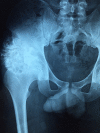
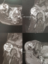
Chondrosarcoma is a rare malignant bone tumor. It can arise de novo or secondary to a malignant transformation of a benign underlying cartilage tumor. Secondary chondrosarcoma arising from solitary benign osteochondroma is extremely rare and data show that the reported incidence of osteochondroma of the pelvis is very low. We here report the case of a 20-year old patient with chondrosarcoma secondary to malignant transformation of an osteochondroma of the right wing of ilium, adjacent to the sacroiliac joint.
Le chondrosarcome est une tumeur osseuse maligne rare. Elle peut être primitive ou secondaire à une transformation maligne d'une tumeur cartilagineuse bénigne sous-jacente. Le chondrosarcome secondaire découlant d'un ostéochondrome solitaire bénin est extrêmement rare et les données montrent que l'incidence rapportée de l'ostéochondrome du bassin est très faible. Nous rapportons le cas d'un patient âgé de 20 ans présentant un chondrosarcome secondaire à la transformation maligne d'un ostéochondrome de l'aile sacro-iliaque droite.
Keywords: Ostéochondrome; chondrosarcome; sacro-iliaque; transformation maligne.
Conflict of interest statement
Les auteurs ne déclarent aucun conflit d'intérêts.
Figures


References
-
- Mark Murphey D, James Choi J, Mark Kransdorf J, Donald Flemming J, Frances Gannon H. Imaging of Osteochondroma: variants and complications with Radiologic-Pathologic Correlation. From the Archives of the AFIP. 2000;20(5):1407–34. - PubMed
-
- Kingsley Chin R, Jaehon Kim M. A rare anterior sacral osteochondroma presenting as sciatica in an adult: a case report and review of the literature. The Spine Journal. 2010;10(5):e1–e4. - PubMed
-
- Lozano Martínez GA, Llauger Rosselló YJ. Condrosarcoma secundario: correlación radiopatológica. Radiología. 2015;57(4):344–359. - PubMed
-
- Moulay Youssef Alaoui Lamrani, Mohammed El Idrissi, Meriem Boubbou, Abdelmajid El Mrini, Mustapha Maâroufi. Les ostéochondromes: aspects clinico-radiologiques, à propos de 12 cas. Pan African Medical Journal. 2016;23:109.
Publication types
MeSH terms
LinkOut - more resources
Full Text Sources
Medical
