. 2019 Jul 2;10(42):4276-4289.
doi: 10.18632/oncotarget.26945.

AGR2, a unique tumor-associated antigen, is a promising candidate for antibody targeting

Affiliations

AGR2, a unique tumor-associated antigen, is a promising candidate for antibody targeting

Alvin Y Liu et al. Oncotarget. .

Abstract

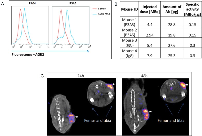
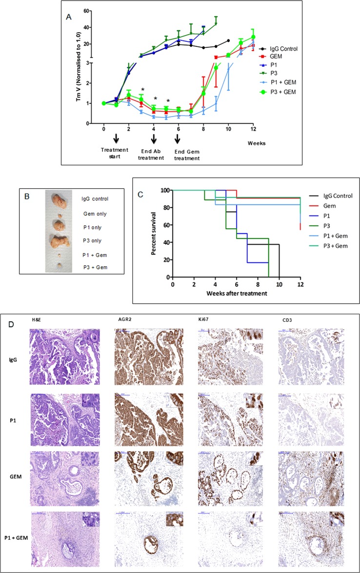
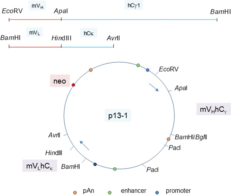
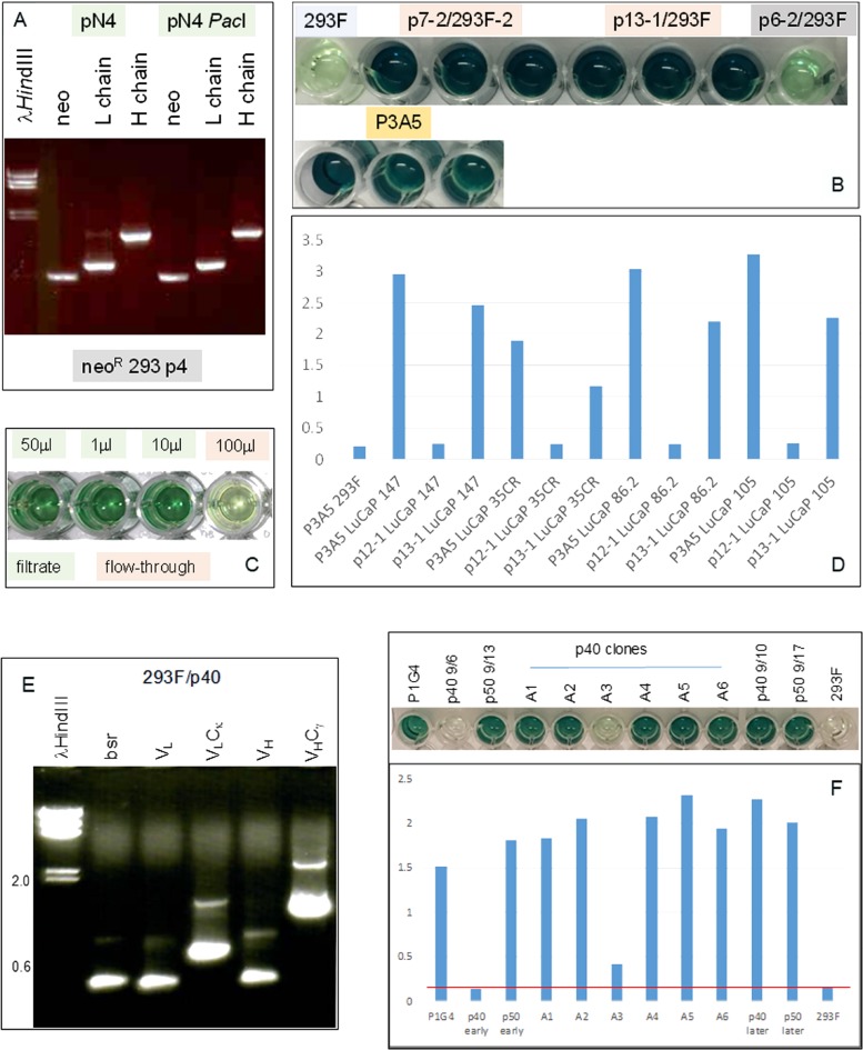
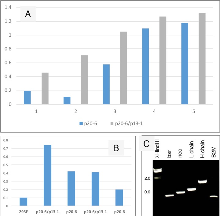
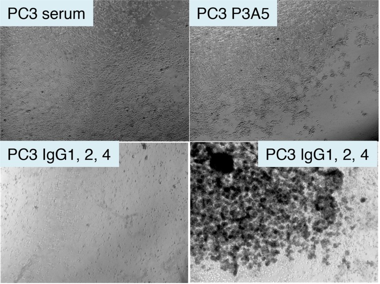
Anterior gradient 2 (AGR2), a protein disulfide isomerase, shows two subcellular localizations: intracellular (iAGR2) and extracellular (eAGR2). In healthy cells that express AGR2, the predominant form is iAGR2, which resides in the endoplasmic reticulum. In contrast, cancer cells secrete and express eAGR2 on the cell surface. We wanted to test if AGR2 is a cancer-specific tumor-associated antigen. We utilized two AGR2 antibodies, P3A5 and P1G4, for in vivo tumor localization and tumor growth inhibition. The monoclonal antibodies recognized both human AGR2 and mouse Agr2. Biodistribution experiments using a syngeneic mouse model showed high uptake of P3A5 AGR2 antibody in xenografted eAgr2+ pancreatic tumors, with limited uptake in normal tissues. In implanted human patient-derived eAGR2+ pancreatic cancer xenografts, tumor growth inhibition was evaluated with antibodies and Gemcitabine (Gem). Inhibition was more potent by P1G4 + Gem combination than Gem alone or P3A5 + Gem. We converted these two antibodies to human:mouse chimeric forms: the constructed P3A5 and P1G4 chimeric mVLhCκ and mVHhCγ (γ1, γ2, γ4) genes were inserted in a single mammalian expression plasmid vector, and transfected into human 293F cells. Expressed human:mouse chimeric IgG1, IgG2 and IgG4 antibodies retained AGR2 binding. Increase in IgG yield by transfected cells could be obtained with serial transfection of vectors with different drug resistance. These chimeric antibodies, when incubated with human blood, effectively lysed eAGR2+ PC3 prostate cancer cells. We have, thus, produced humanized anti-AGR2 antibodies that, after further testing, might be suitable for treatment against a variety of eAGR2+ solid tumors.

Keywords: chimeric antibody; eAGR2; pancreatic cancer; prostate cancer; tumor localization.


Conflict of interest statement

CONFLICTS OF INTEREST The authors do not have any conflicts of interest.

Figures

Figure 1
Figure 1. Cell surface expression of AGR2.
(A) Fluorescence shifts of P1G4- and P3A5-labeled DT6606 primary pancreatic cancer cells (vs. control IgG) indicate that eAgr2 is present on the cell surface. Two cytograms are shown. (B) The table shows labeling of antibodies. (C) The representative images show DT6606 tumor uptake of radiolabeled P3A5 (marked by *) at 24 h and 48 h post-injection. No significant label could be detected in iAgr2-positive normal tissues.
Figure 2
Figure 2. Tumor growth inhibition by drug in combination with antibodies.
(A) PDX tumor growth in mice in response to treatment with ɑ-AGR2 P1 (P1G4) and P3 (P3A5), alone or in combination with Gem demonstrate that antibodies alone produced minimal effect; the best tumor suppression was achieved in the P1 + Gem group. Tumor volume is indicated on the y-axis, and treatment duration in weeks on the x-axis. Differences in tumor volumes between P1 + Gem vs. Gem were statistically significant (*) at weeks 3-5. (B) Tumors extracted at week 6 from the different groups are compared. (C) The Kaplan–Meier plot shows the percentage survival (y-axis) of mice across all six groups (in weeks on the x-axis). (D) Representative immunohistochemistry images on tumor tissue samples from IgG control, P1, Gem and P1 + Gem confirm expression of AGR2 in all tumors. Ki67 staining indicated that the tumors treated with Gem still had high proliferation rate, which was limited in P1 + Gem-treated tumors. The tumors from Gem- and P1 + Gem-treated mice exhibited abundant CD3+ T cell infiltration. Size bars = 200 μm. Inserts at the top right of individual images are at 200x magnification.
Figure 3
Figure 3. Plasmid vector for P3A5 H and L chain expression.
The joined mouse VH module (EcoRV-ApaI) and human Cγ1 module (ApaI-BamHI), as well as the joined mouse VL module (BamHI-HindIII) and human Cκ module (HindIII-AvrII) are shown. The H and L chain genes are cloned in the orientation depicted in plasmid p13-1. Gene expression control elements are identified. PacI is used to linearize the plasmid for transfection. Plasmid p20-6 has bsr instead of neo. Plasmids p40-1 and p50-1 (not shown) are the corresponding neo and bsr vectors for chimeric P1G4.
Figure 4
Figure 4. Immunoglobulin expression in transfected 293F cells.
(A) RT-PCR analysis for neo (560 bp), L (720 bp) and H (1420 bp) chains in cells transfected with plasmid pN4 at passage 4 (p4) to show that the transgenes were being transcribed at equivalent levels. (B) ELISA result shows AGR2 binding by IgG1 from transfected 293F cells (in triplicate). The color intensity indicates similar binding affinity between the IgG1 constructs p7-2 and p13-1 vs. P3A5. Background consists of untransfected 293F and p6-2, which is a non-productive construct. Detection is by either HRP anti-human IgG for the transfected cell media or HRP anti-mouse IgG2a for P3A5. (C) ELISA shows IgG isolation by spin filtration. Background level was detected in the 100 μL flow-through well (4th) vs. the three wells (1st – 3rd) containing different amounts of the filtrate/retentate. (D) Histogram representation of ELISA data shows comparable binding between 293F-synthesized human IgG and P3A5 in tissue digestion media of LuCaP lines (147, 35CR, 86.2, 105) with different levels of AGR2 expression. p12-1 is a defective construct, and served as negative control. Absorbance units are indicated on the y-axis. (E) RT-PCR analysis of 293F/p40-1 P1G4 chimeric detected transcripts for bsr (420 bp), VL, VLCκ, VH, and VHCγ1. (F) The histogram bars show AGR2 binding activities detected in media supernatant of p40, p50, and p40 clones A1-A6 (absorbance units on the y-axis). The positive control is mouse P1G4 and the negative control 293F cells. “Early” and “later” indicate media harvested during early and late passages, respectively, of cultures after drug selection.
Figure 5
Figure 5. Increased IgG production from serially transfected 293F cells.
(A) Cell-free media supernatant from increasing amounts of cells seeded in culture wells were assayed for AGR2-binding activity. The source of AGR2 was provided by LNCaP cells transfected with AGR2 (based on the same plasmid vector). Absorbance units are shown on the y-axis. (B) The volumes of media supernatant obtained from similar numbers of cells were assayed. The negative control is media from untransfected 293F cells. Absorbance units are shown on the y-axis. (C) Transcript levels of bsr, neo, L and H chains, B2M were detected in the doubly transfected clone A3.
Figure 6
Figure 6. Treatment of AGR2+ PC3 cells by chimeric antibodies.
Shown are photomicrographs of plated eAGR2+ PC3 cells incubated in human serum (top left); serum with P3A5 (top right); serum with IgG1, IgG2, IgG4 (bottom left and right).

Similar articles

Cited by

References

    1. Pascal LE, Vêncio RZ, Page LS, Liebeskind ES, Shadle CP, Troisch P, Marzolf B, True LD, Hood LE, Liu AY. Gene expression relationship between prostate cancer cells of Gleason 3, 4 and normal epithelial cells as revealed by cell type-specific transcriptomes. BMC Cancer. 2009; 9:452. 10.1186/1471-2407-9-452. . - DOI - PMC - PubMed
    1. Ramachandran V, Arumugam T, Wang H, Logsdon CD. Anterior gradient 2 is expressed and secreted during the development of pancreatic cancer and promotes cancer cell survival. Cancer Res. 2008; 68:7811-7818. 10.1158/0008-5472.CAN-08-1320. . - DOI - PMC - PubMed
    1. Dumartin L, Whiteman HJ, Weeks ME, Hariharan D, Dmitrovic B, Iacobuzio-Donahue CA, Brentnall TA, Bronner MP, Feakins RM, Timms JF, Brennan C, Lemoine NR, Crnogorac-Jurcevic T. AGR2 is a novel surface antigen that promotes the dissemination of pancreatic cancer cells through regulation of cathepsins B and D. Cancer Res. 2011; 71:7091-7102. 10.1158/0008-5472.CAN-11-1367. . - DOI - PMC - PubMed
    1. Chen YT, Ho CL, Chen PK, Chen YL, Chang CF. Anterior gradient 2: a novel sensitive tumor marker for metastatic oral cancer. Cancer Lett. 2013; 339:270-278. 10.1016/j.canlet.2013.06.025. . - DOI - PubMed
    1. Fritzsche FR, Dahl E, Pahl S, Burkhardt M, Luo J, Mayordomo E, Gansukh T, Dankof A, Knuechel R, Denkert C, Winzer KJ, Dietel M, Kristiansen G. Prognostic relevance of AGR2 expression in breast cancer. Clin Cancer Res. 2006; 12:1728-1734. 10.1158/1078-0432.CCR-05-2057. . - DOI - PubMed