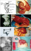
Congenital Segmental Intestinal Dilatation: A 25-Year Review with Long-Term Follow-up at the Medical University of Innsbruck, Austria
- PMID: 31304051
- PMCID: PMC6624109
- DOI: 10.1055/s-0039-1693164
Congenital Segmental Intestinal Dilatation: A 25-Year Review with Long-Term Follow-up at the Medical University of Innsbruck, Austria
Abstract
Background and Aim Congenital segmental intestinal dilatation (CSID) is a neonatal condition with unclear etiology and pathogenesis. Typically, the newborn with CSID presents with a limited (circumscribed) bowel dilatation, an abrupt transition between normal and dilated segments, neither intrinsic nor extrinsic perilesional obstruction, and no aganglionosis or neuronal intestinal dysplasia. We aimed to review this disease and the long-term follow-up at the Children's Hospital of the Medical University of Innsbruck, Tyrol, Austria. Study Design Retrospective 25-year review of medical charts, electronic files, and histopathology of neonates with CSID. Results We identified four infants (three girls and one boy) with CSID. The affected areas included duodenum, ileum, ascending colon, and sigmoid colon. Noteworthy, all patients presented with a cardiovascular defect, of which two required multiple cardiac surgical interventions. Three out of the four patients recovered completely. To date, the three infants are alive. Conclusion This is the first report of patients with CSID and cardiovascular defects. The clinical and surgical intervention for CSID also requires a thorough cardiologic evaluation in these patients. CSID remains an enigmatic entity pointing to the need for joint forces in identifying common loci for genetic investigations.
Keywords: Hirschsprung's disease; aganglionosis; dilatation; heart defect; intestine; obstruction.
Conflict of interest statement
Figures


Similar articles
-
Segmental aganglionosis in Hirschsprung's disease in newborns - a case report.Rom J Morphol Embryol. 2015;56(2):533-6. Rom J Morphol Embryol. 2015. PMID: 26193224
-
[Segmental dilatation of the ileum].An Esp Pediatr. 1984 Dec;21(9):847-51. An Esp Pediatr. 1984. PMID: 6529043 Spanish.
-
Segmental dilatation of intestine presenting as partial intestinal obstruction in a child.APSP J Case Rep. 2014 May 21;5(2):19. eCollection 2014 May. APSP J Case Rep. 2014. PMID: 25057472 Free PMC article.
-
Congenital aganglionic megacolon in Nigerian adults: two case reports and review of the literature.Niger J Clin Pract. 2011 Apr-Jun;14(2):249-52. doi: 10.4103/1119-3077.84032. Niger J Clin Pract. 2011. PMID: 21860150 Review.
-
Unusual presentation of sacrococcygeal teratomas and associated malformations in children: clinical experience and review of the literature.Ann Ital Chir. 2013 May-Jun;84(3):333-46. Ann Ital Chir. 2013. PMID: 23160138 Review.
Cited by
-
Pediatrics: An Evolving Concept for the 21st Century.Diagnostics (Basel). 2019 Nov 25;9(4):201. doi: 10.3390/diagnostics9040201. Diagnostics (Basel). 2019. PMID: 31775294 Free PMC article.
-
Congenital segmental dilatation of the colon in a newborn: a radiologic perspective of an unusual entity.Pediatr Radiol. 2023 Jul;53(8):1722-1725. doi: 10.1007/s00247-023-05639-0. Epub 2023 Mar 8. Pediatr Radiol. 2023. PMID: 36884051
-
Congenital Segmental Dilatation of Ascending Colon with Distal Microcolon: A Diagnostic Dilemma.J Indian Assoc Pediatr Surg. 2023 Nov-Dec;28(6):529-531. doi: 10.4103/jiaps.jiaps_132_23. Epub 2023 Nov 2. J Indian Assoc Pediatr Surg. 2023. PMID: 38173636 Free PMC article.
-
Congenital segmental dilatation of the intestine in a neonate.BMJ Case Rep. 2023 Dec 28;16(12):e256842. doi: 10.1136/bcr-2023-256842. BMJ Case Rep. 2023. PMID: 38154863
References
-
- Ben Brahim M, Belghith M, Mekki M et al.Segmental dilatation of the intestine. J Pediatr Surg. 2006;41(06):1130–1133. - PubMed
-
- Takawira C, D'Agostini S, Shenouda S, Persad R, Sergi C. Laboratory procedures update on Hirschsprung disease. J Pediatr Gastroenterol Nutr. 2015;60(05):598–605. - PubMed
-
- Burtelow M A, Longacre T A. Utility of microtubule associated protein-2 (MAP-2) immunohistochemistry for identification of ganglion cells in paraffin-embedded rectal suction biopsies. Am J Surg Pathol. 2009;33(07):1025–1030. - PubMed
-
- Chisholm K M, Longacre T A. Utility of peripherin versus MAP-2 and calretinin in the evaluation of Hirschsprung disease. Appl Immunohistochem Mol Morphol. 2016;24(09):627–632. - PubMed
-
- Yang W I, Oh J T. Calretinin and microtubule-associated protein-2 (MAP-2) immunohistochemistry in the diagnosis of Hirschsprung's disease. J Pediatr Surg. 2013;48(10):2112–2117. - PubMed
Publication types
LinkOut - more resources
Full Text Sources

