

doi: 10.1093/ofid/ofz285.
eCollection 2019 Jul.
A Fatal Case of Disseminated Microsporidiosis Due to Anncaliia algerae in a Renal and Pancreas Allograft Recipient
Affiliations
- PMID: 31304191
- PMCID: PMC6612885
- DOI: 10.1093/ofid/ofz285
Item in Clipboard
A Fatal Case of Disseminated Microsporidiosis Due to Anncaliia algerae in a Renal and Pancreas Allograft Recipient
Open Forum Infect Dis.
.
Abstract
Microsporidiosis is an emerging opportunistic infection in immunocompromised patients. We report a case of fatal disseminated Anncaliia algerae infection in a profoundly immunosuppressed pancreas and kidney transplant recipient.
Keywords: Brachiola; Nosema; immunocompromised; microspo-ridiosis; microsporidia; opportunistic.
Figures
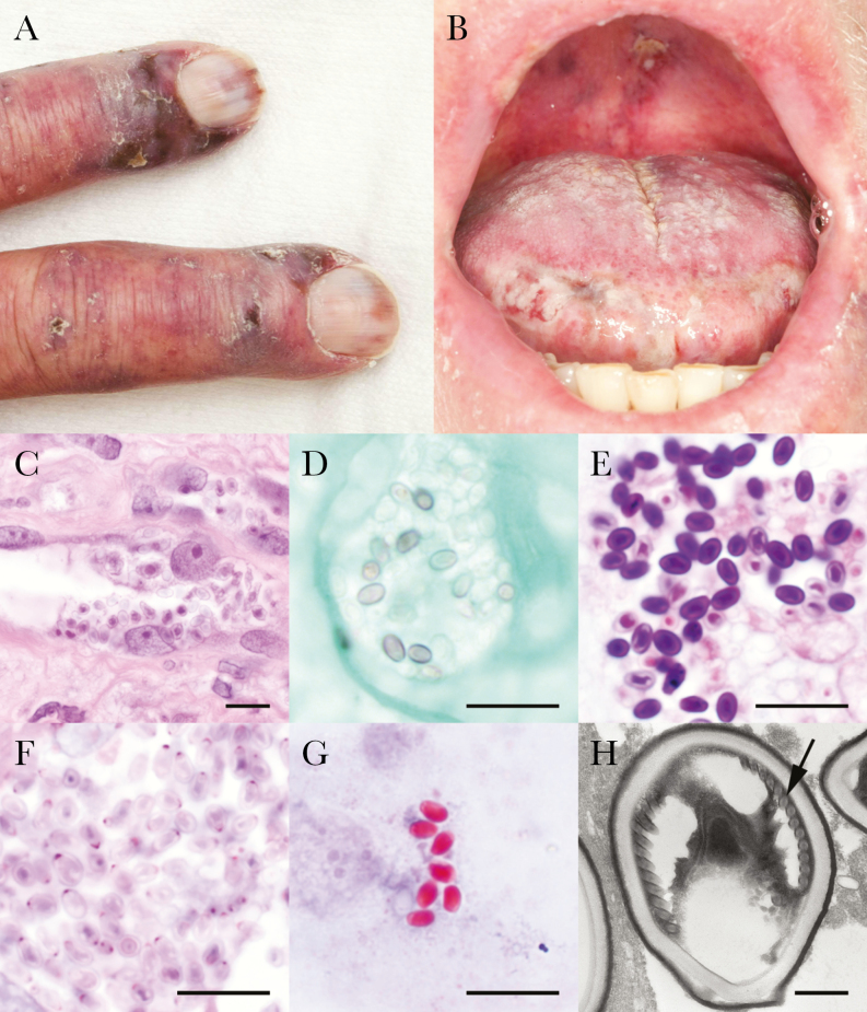
The patient developed several clinical manifestations of disseminated microsporidiosis due to Anncaliia algerae, including (A) ulcerative fingers and (B) oral lesions, as well as ulcerative intranasal lesions and a diffuse papular rash of the lower extremities (not shown). By light microscopic examination, spores were visible using (C) routine hematoxylin and eosin staining, (D) focally positive by Gomori methenamine silver stain, and (E) strongly Gram-positive by Gram stain. (F) The Periodic acid–Schiff stain showed polar dot-like positivity, which is characteristic of microsporidial spores. Spores also were stained bright red using (G) the Ryan blue modified trichrome stain for microsporidia. (H) Electron microscopy showed features of a Nosema-like microsporidia, including large size, a thick cell wall, and a single row of polar filaments (arrow). Scale bars represent 20 µm, except in (H) in which the scale bar is 500 nm.
References
-
- Kester KE, Turiansky GW, McEvoy PL. Nodular cutaneous microsporidiosis in a patient with AIDS and successful treatment with long-term oral clindamycin therapy. Ann Intern Med 1998; 128:911–4. - PubMed
-
- Weber R, Deplazes P, Mathis A. Microsporidia. In: Jorgensen J, Pfaller M, Carroll K, Funke G, Landry M, Richter S, Warnock D, eds. Manual of Clinical Microbiology. 11th ed. Washington, DC: ASM Press; 2015:2209–19.
LinkOut - more resources
Full Text Sources
