. 2017 Oct 30:1:3.
doi: 10.1038/s41538-017-0003-3. eCollection 2017.

Direct interaction of food derived colloidal micro/nano-particles with oral macrophages

Affiliations

Direct interaction of food derived colloidal micro/nano-particles with oral macrophages

Lijing Ke et al. NPJ Sci Food. .

Abstract

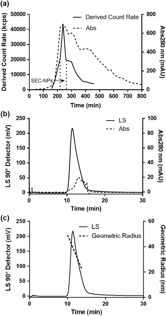
Like any typical food system, bone soup (or broth), a traditional nourishing food in many cultures, contains a colloid dispersion of self-assembled micro/nano-particles. Food ingestion results in the direct contact of food colloidal MNPs with immune cells. Will they ever interact with each other? To answer the question, MNPs and NPs were separated from porcine bone soup and labeled with Nile Red, and their uptake by murine oral macrophages and its consequent effects were investigated. Colloidal particle samples of UF-MNPs and SEC-NP were prepared from porcine bone soup by ultrafiltration (UF) and size-exclusion chromatography, respectively. Their mean hydrodynamic diameters were 248 ± 10 nm and 170 ± 1 nm with dominant composition of protein and lipid. Particles in both samples were found to be internalized by oral macrophages upon co-incubation at particle/cell ratios of 14,000/1. In normal oral macrophages, the particle uptake exerted influence neither on the cellular cytosolic membrane potential (V mem) nor mitochondrial superoxide level, as were indicated with fluorescent dyes of DiBAC4(3) and MitoSOX Red, respectively. However, when oral macrophages were challenged by peroxyl radical inducer AAPH, the engulfment of UF-MNPs and SEC-NPs mitigated the peroxyl radical induced membrane hyperpolarization effect by up to 70%, and the suppression on the oxygen respiration in mitochondria by up to 100%. Those results provide evidence of the direct interaction between food colloidal particles with immune cells, implying a possible new mode of food-body interaction.

Keywords: Mucosal immunology; Nanoparticles.


Conflict of interest statement

Competing interestsThe authors declare that they have no competing financial interests.

Figures

Fig. 1
Fig. 1
SEC chromatographic isolation of SEC-NPs from porcine bone soup. Bone soup was fractioned by a Sephacryl S-1000 SF column (1.0 × 100 cm) equilibrated with 0.02 M phosphate buffer (pH 7.4). The column was eluted with the same buffer at a flow rate of 0.50 mL/min at 40 °C with UV and Zetasizer Nano-ZS detector (Malven, UK). The purity and molar mass of SEC-NPs was determined by a SEC-MALLS (Size-Exclusion HPLC coupled with Multi-Angle Laser Light Scattering). A TSK gel G6000PWxl HPLC column (0.78 × 300 cm, Tosoh Bioscience, Japan) was equilibrated and eluted with phosphate buffer (0.1 M, pH7.4) at flow rate 0.80 mL/min. a: SEC chromatogram (Sephacryl S-1000 SF column) of the porcine bone soup is presented as the Derived Count Rate (solid line) and UV absorptance at 280 nm (dash line). The fraction of NPs was collected and named SEC-NPs, as indicated by two dot line. b: The SEC-MALLS chromatogram (TSK gel G6000PWxl HPLC column) of SEC-NPs fraction is presented as the light scattering intensity at 90° of MALLS (solid line) and UV absorptance at 280 nm (dash line). c: The solid line is the SEC-MALLS chromatogram wherein the dash line represents the geometric radius distributions (22–49 nm) of the light scattering peak
Fig. 2
Fig. 2
TEM and Cryo-TEM micrographs of food derived micro/nano-particles TEM images: magnification × 10000, scale bar “▬” as 200 nm; a: porcine bone soup MNPs; b: UF-MNPs isolated from porcine bone soup; c: SEC-NPs isolated from porcine bone soup. Cryo-TEM images: magnification × 8000; d: porcine bone soup MNPs, scale bar “▬” as 500 nm; e: UF-MNPs isolated from porcine bone soup, scale bar “▬” as 500 nm; f: SEC-NPs isolated from porcine bone soup, scale bar “▬” as 500 nm. Green arrows: particles with diameter of 40~100 nm; Red arrows: particles with diameter of around 250 nm
Fig. 3
Fig. 3
Fluorescent microscopic graph of food derived micro/nano- particles a: fluorescence micrograph of Nile Red stained UF-MNPs from porcine bone soup; b and c: the hydrodynamic diameter and ζ-potential of SEC-NPs, isolated from porcine bone soup, wherein no significant difference was observed before and after Nile red staining
Fig. 4
Fig. 4
Uptake of food derived micro/nano-particles via oral mucosal macrophages. The red dots observed in cellular plasma (indicated with white or black arrows on the graphs) are Nile-red stained porcine bone soup UF-MNPs and SEC-NPs. The cell nuclei were stained in blue with a fluorescent dye Hoechst 33342. af: confocal fluorescence micrographs of macrophages, the particle/cell ratios were 3500/1 on oral macrophages. Cell images at different magnifications are given wherein the images of a single cell (framed with white square in diagram a, b, and c) are enlarged and presented as diagram df, scale bar = 5 μm. The red and blue fluorescent cell images are merged and presented on the left, the fluorescent and bright cell images are merged and presented on the right
Fig. 5
Fig. 5
Effects of food derived micro/nano-particles on cell plasma membrane potential (indicated with green fluorescence of DiBAC4(3)) and mitochondrial superoxide (indicated with red fluorescence of MitoSOX) of murine oral mucosal macrophages. Cells were loaded with 3 μM MitoSOX Red for 10 min and then 2.5 μM DiBAC4(3) for 15 min at 37 °C. For AAPH treated cells (6.4 μM, incubated for 120 min), they were loaded with fluorescence dyes prior to the addition of AAPH and micro/nano-particles. a: Fluorescent micrographs (red & green fluorescence, taken by a Leica DMI3000 B Inverted Microscope, excitation at 515~560 nm and 450~490 nm, respectively) of loaded cells, merged with software LAS (version 4.2.0) wherein UF-MNPs/cell ratio = 3500/1,700/1 and 140/1; SEC-NPs/cell ratio = 3500/1,700/1 and 140/1. b: V mem-dependent fluorescence traces (1 data-point/10 min for 120 min, excitation/emission at 493/516 nm for DiBAC4(3)) of loaded macrophages in a 96-well microplate were measured simultaneously by a fluorometric plate reader (Flexstation3, Molecular Devices, USA). c: the diagram shows the impacts of UF-MNPs, SEC-NPs and AAPH on the mitochondrial superoxide level of macrophages, indicated by MitoSOX Red fluorescence (excitation/emission at 510/580 nm). *: P < 0.05 v.s. Control, **: P < 0.01 v.s. Control; #: P < 0.05 v.s. AAPH, ##: P < 0.01 v.s. AAPH. The oral macrophages partially presented green-yellowish color due to the overlap between two fluorescent probes in cell compartments or cell groups. Among normal macrophages, cells with higher level of mitochondrial superoxide (brighter red fluorescence) tend to have hyperpolarized membranes (weaker green fluorescence) which carry more intensively the negative charge. AAPH induced peroxyl radicals eliminated the DiBAC4(3) fluorescence as well as decreased MitoSOX Red fluorescence, indicating the hyperpolarization of cell membrane and down-regulated mitochondrial oxygen respiratory

References

    1. Shan W, et al. Overcoming the diffusion barrier of mucus and absorption barrier of epithelium by self-assembled nanoparticles for oral delivery of insulin. ACS Nano. 2015;9:2345–2356. doi: 10.1021/acsnano.5b00028. - DOI - PubMed
    1. Wang YJ, et al. Tea nanoparticle, a safe and biocompatible nanocarrier, greatly potentiates the anticancer activity of doxorubicin. Oncotarget. 2016;7:5877–5891. doi: 10.18632/oncotarget.6711. - DOI - PMC - PubMed
    1. Penalva R, et al. Casein nanoparticles as carriers for the oral delivery of folic acid. Food Hydrocoll. 2015;44:399–406. doi: 10.1016/j.foodhyd.2014.10.004. - DOI
    1. Zimet P, Rosenberg D, Livney YD. Re-assembled casein micelles and casein nanoparticles as nano-vehicles for??-3 polyunsaturated fatty acids. Food Hydrocoll. 2011;25:1270–1276. doi: 10.1016/j.foodhyd.2010.11.025. - DOI
    1. Shutava TG, et al. Layer-by-layer-coated gelatin nanoparticles as a vehicle for delivery of natural polyphenols. ACS Nano. 2009;3:1877–1885. doi: 10.1021/nn900451a. - DOI - PubMed