Review
. 2019 Jun 27:4:50.
doi: 10.21037/tgh.2019.06.02. eCollection 2019.

Pathology of pancreatic cancer

Affiliations
Review

Pathology of pancreatic cancer

Lena Haeberle et al. Transl Gastroenterol Hepatol. .

Abstract

Pancreatic ductal adenocarcinoma (PDAC) is a highly aggressive malignancy and estimated to become the second leading cause of cancer-related deaths by 2030. Although overall 5-year survival rates have constantly remained below 10% for the last decades, several key points important for accurate patient stratification have emerged during recent years. These key points include a highly standardized gross examination of PDAC resection specimens, using an axial slicing technique and inking of the circumferential resection margin (CRM), as well as a meticulous microscopic examination, taking into account the prognostic relevance of factors such as the exact resection status (R0 vs. R1 1-mm vs. R1 resection), histopathological tumor grading and the so-called lymph node ratio (LNR). With increasing use of neoadjuvant therapy in PDAC, tumor regression grading (TRG) for PDAC is currently rising in relevance in order to stratify and manage pre-operatively treated PDAC patients. As all current TRG systems for PDAC are unsatisfactory, new standardized international protocols are urgently needed. Several morphological subtypes of PDAC exist, some of which share the same molecular background with classical PDAC, while others are characterized by a distinct molecular pathogenesis. While some show a prognosis similar to classical PDAC, other subtypes stand out due to a better or even worse prognosis than classical PDAC. Prognostic relevant molecular subtypes of PDAC have been proposed as well, however, limitations of used cohorts and the lacking correlation of molecular subtypes with histomorphological subtypes limit the translation of these findings into valuable clinical applications. Lastly, several macroscopic and microscopic precursor lesions of PDAC have been described in genetically engineered mouse models (GEMM) and humans in recent times, providing further insight into PDAC carcinogenesis. In addition, improved diagnosis of PDAC precursors represents a chance to select patients for resection before invasive PDAC is present.

Keywords: Histomorphological variants; molecular subtypes; neoadjuvant treatment; pancreatic ductal adenocarcinoma (PDAC); precursor lesions; standardized pathology report.


Conflict of interest statement

Conflicts of Interest: The authors have no conflicts of interest to declare.

Figures

Figure 1
Figure 1
Gross morphology and histomorphology of PDAC. (A) Gross morphology of PDAC after axial slicing. PDAC presents as solid ill-defined white-yellowish mass of the pancreas head (circle). The ventral CRM was inked blue (black arrow), the medial CRM green (white arrow) and the posterior CRM black (arrowhead). (B) Histomorphology of PDAC. Tumor cells are present within 1 mm of the ventral CRM, highlighted with green ink, warranting the diagnosis of “R1 1-mm” status. (C) Typical histomorphology of PDAC. Small to medium-sized irregular glands are embedded in a desmoplastic stroma. (D) Histomorphology of PDAC after neoadjuvant therapy. Tumor cells show signs of regression, such as vacuolization of the cytoplasm and marked nuclear atypia, although the distinction between regressive changes and pre-existent tumor cell features is nearly impossible. (B) 100×, H&E; (C) 100×, H&E; (D) 100×, H&E. PDAC, pancreatic ductal adenocarcinoma; CRM, circumferential resection margin.
Figure 2
Figure 2
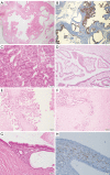
Histomorphologic variants of PDAC. (A) Histomorphology of adenosquamous pancreatic carcinoma, consisting of a glandular and a solid-squamous tumor component; (B) immunohistochemistry of adenosquamous pancreatic carcinoma. Squamous component stains positive for p40; (C) histomorphology of anaplastic pancreatic carcinoma displaying perineural invasion by dispersed, highly pleomorphic tumor cells; (D) immunohistochemistry of anaplastic pancreatic carcinoma with positivity for Vimentin; (E) histomorphology of colloid (mucinous) pancreatic carcinoma with invasive tumor cells embedded in extensive mucin pools; (F) PAS-positivity of mucin pools in colloid (mucinous) pancreatic carcinoma. (A) 100×, H&E; (B) 100×, p40; (C) 400×, H&E; (D) 200×, Vimentin; (E) 100×, H&E; (F) 20×, PAS.PDAC, pancreatic ductal adenocarcinoma.
Figure 3
Figure 3
Macroscopic precursor lesions of PDAC. (A) Histomorphology of IPMN gastric type, low grade, displaying intraductal-papillary epithelial proliferations resembling gastric foveolae; (B) MUC5 expression in IPMN gastric type, low grade; (C) histomorphology of IPMN gastric type, high grade, characterized by a more solid growth and larger, pleomorphic nuclei; (D) histomorphology of IPMN intestinal type, high grade, with long finger-like papillae resembling villous adenoma of the colon; (E) histomorphology of IPMN pancreatobiliary type, high grade, consisting of complex arborizing papillae; (F) IPMN pancreatobiliary type, high grade, with associated invasive carcinoma (lower right); (G) histomorphology of MCN, low grade, of the pancreas, showing columnar mucinous epithelium and characteristic subepithelial “ovarian-like” stroma with high cellularity; (H) expression of oestrogen receptor in the “ovarian-like” stroma of MCN. (A) 20×, H&E; (B) 20×, MUC5; (C) 200×, H&E; (D) 100×, H&E; (E) 100×, H&E; (F) 100×, H&E; (G) 200×, H&E; (F) 400×, oestrogen receptor. PDAC, pancreatic ductal adenocarcinoma; IPMN, intraductal papillary mucinous neoplasms.

Similar articles

Cited by

References

    1. Cascinu S, Falconi M, Valentini V, et al. Pancreatic cancer: ESMO Clinical Practice Guidelines for diagnosis, treatment and follow-up. Ann Oncol 2010;21 Suppl 5:v55-58. 10.1093/annonc/mdq165 - DOI - PubMed
    1. Peixoto RD, Speers C, McGahan CE, et al. Prognostic factors and sites of metastasis in unresectable locally advanced pancreatic cancer. Cancer Med 2015;4:1171-7. 10.1002/cam4.459 - DOI - PMC - PubMed
    1. Esposito I, Kleeff J, Bergmann F, et al. Most pancreatic cancer resections are R1 resections. Ann Surg Oncol 2008;15:1651-60. 10.1245/s10434-008-9839-8 - DOI - PubMed
    1. Verbeke CS, Leitch D, Menon KV, et al. Anthoney, Redefining the R1 resection in pancreatic cancer. Br J Surg 2006;93:1232-7. 10.1002/bjs.5397 - DOI - PubMed
    1. Menon KV, Gomez D, Smith AM, et al. Impact of margin status on survival following pancreatoduodenectomy for cancer: the Leeds Pathology Protocol (LEEPP). HPB 2009;11:18-24. 10.1111/j.1477-2574.2008.00013.x - DOI - PMC - PubMed