Review
. 2019 Jul 15;6(1):23.
doi: 10.1186/s40580-019-0193-2.

Current trends and challenges in cancer management and therapy using designer nanomaterials

Affiliations
Review

Current trends and challenges in cancer management and therapy using designer nanomaterials

P N Navya et al. Nano Converg. .

Abstract

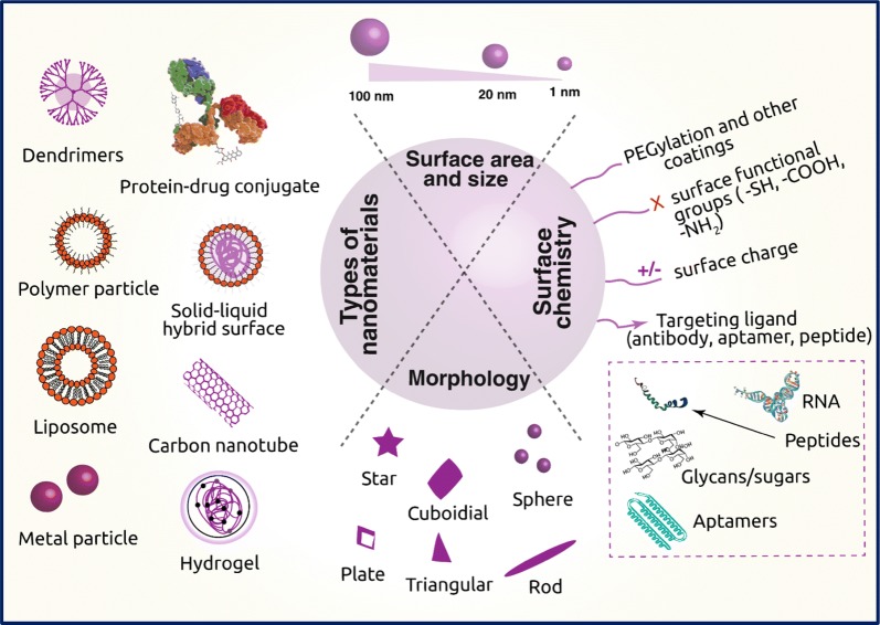
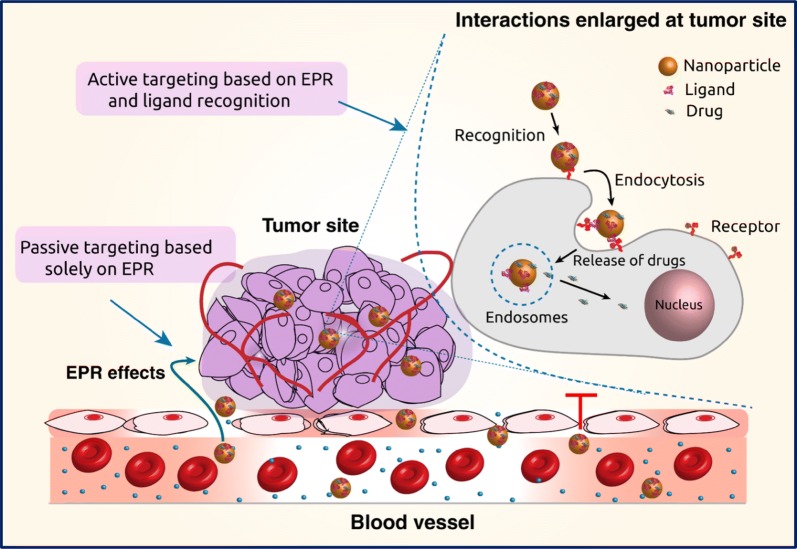
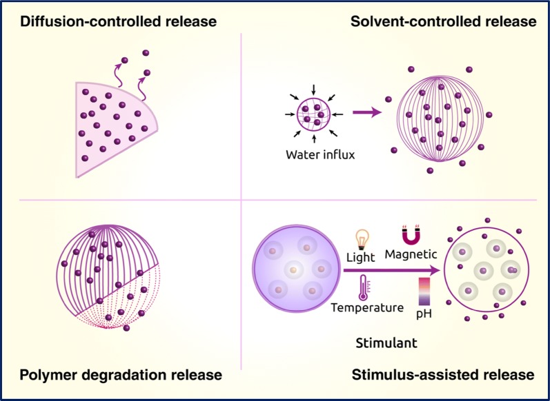
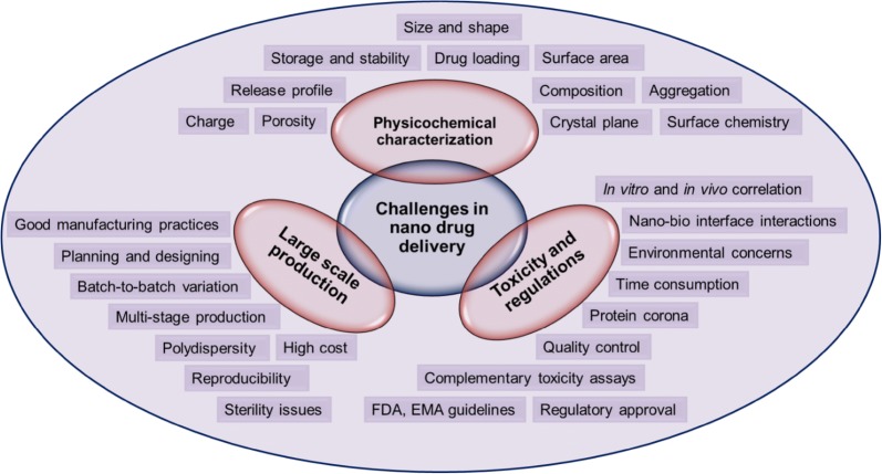
Nanotechnology has the potential to circumvent several drawbacks of conventional therapeutic formulations. In fact, significant strides have been made towards the application of engineered nanomaterials for the treatment of cancer with high specificity, sensitivity and efficacy. Tailor-made nanomaterials functionalized with specific ligands can target cancer cells in a predictable manner and deliver encapsulated payloads effectively. Moreover, nanomaterials can also be designed for increased drug loading, improved half-life in the body, controlled release, and selective distribution by modifying their composition, size, morphology, and surface chemistry. To date, polymeric nanomaterials, metallic nanoparticles, carbon-based materials, liposomes, and dendrimers have been developed as smart drug delivery systems for cancer treatment, demonstrating enhanced pharmacokinetic and pharmacodynamic profiles over conventional formulations due to their nanoscale size and unique physicochemical characteristics. The data present in the literature suggest that nanotechnology will provide next-generation platforms for cancer management and anticancer therapy. Therefore, in this critical review, we summarize a range of nanomaterials which are currently being employed for anticancer therapies and discuss the fundamental role of their physicochemical properties in cancer management. We further elaborate on the topical progress made to date toward nanomaterial engineering for cancer therapy, including current strategies for drug targeting and release for efficient cancer administration. We also discuss issues of nanotoxicity, which is an often-neglected feature of nanotechnology. Finally, we attempt to summarize the current challenges in nanotherapeutics and provide an outlook on the future of this important field.

Keywords: Cancer therapy; Drug delivery; Engineered nanomaterials; Nanotoxicity; Next-generation.


Conflict of interest statement

The authors declare that they have no competing interests.

Figures

Fig. 1
Fig. 1
Schematic representation of different types of nanomaterials employed in cancer therapy, their important physical properties and surface chemistry required to carry drugs
Fig. 2
Fig. 2
Graphical illustration of passive and active drug targeting strategies. In passive targeting, the nanocarriers pass through the leaky walls and accumulate at the tumor site by the enhanced permeability and retention (EPR) effect. Active targeting can be achieved using specific ligands that bind to the receptors on the tumor cells
Fig. 3
Fig. 3
In vitro and in vivo effects of IGF1-IONPs (insulin-like growth factor 1-iron oxide nanoparticles) and IGF1-IONPs-doxorubicin on cell proliferation and viability. a The effect of IGF1R in MIAPaCa-2 cells was assessed by immunofluorescence labeling employing an anti-IGF1R antibody (shown in red color). b Prussian blue staining of cells incubated for 4 h with different treatments at 20 μg/mL of iron equivalent dose. The cells are also counterstained with nuclear fast red. c The in vitro influence of IGF1 and IGF1-IONPs on cell proliferation. The % of viable cells after 96 h incubation with IGF1 or IGF1-IONPs, and for 4 h at equivalent IGF1 concentrations was estimated by cell proliferation assay, wherein *P < 0.05; **P < 0.001. d The in vivo effect on tumor cell proliferation of IGF1-IONPs in human pancreatic PDX-tumor xenografts. By using immunofluorescence labeling of an anti-Ki67 antibody, the Ki67-positive cells in tumor sections after two tail vein injections of 20 mg/kg iron dose of IGF1-IONPs are measured. e In vitro cytotoxicity of unconjugated and conjugated doxorubicin in MIA PaCa-2 cells. The scale bars are 100 μm (adapted with permission from [48])
Fig. 4
Fig. 4
Schematic depiction of diffusion-, solvent-controlled, polymer degradation, and other stimuli reliant drug release
Fig. 5
Fig. 5
Cellular uptake of gold nanoconstructs by U87 glioblastoma cells. A Transmission electron micrographs of Au nanoparticles displaying 13 nm spheres, 50 nm spheres and 40 nm stars; B cellular uptake kinetics of Au nanoparticles-siRNA constructs by cells showing size and shape dependent uptake; C transmission electron images illustrating the process of cellular uptake after treatment with 0.5 nM of Au nanoparticles-siRNA constructs for 24 h. The vesicle membranes disrupted by the treatment with 50 nm spheres is signified by orange arrows, and the nanoconstructs distributed outside the vesicles is represented by yellow arrows (reproduced with permission from [103])
Fig. 6
Fig. 6
Effect of OVA-iron oxide nanoparticles: macrophages activation with different concentrations of OVA, and production of a TNF-α, b IL-6, c IFN-γ. Saline and LPS served as negative and positive control; d size of the tumor measured after 22nd day of mice immunization; e histological sections of different organs on 23rd day after immunization of mice with different treatments (1) control, (2) soluble OVA, (3) iron oxide nanoparticles and (4) OVA-iron oxide nanoparticles (reproduced with permission from [188])
Fig. 7
Fig. 7
Scheme representing the formulation of doxorubicin loaded PEGylated liposome, and doxorubicin loaded lactoferrin modified PEGylated liposome (a); effect of cell viability of free DOX and the liposomal formulations evaluated by MTT assay in HepG2, BEL7402, and SMMC7721 cells at different time intervals (b); relative tumor volume of various liposomal formulations injected to tumor-bearing mice through tail veins every 7 days at a dose of 5 mg/kg DOX (c); change in the body weight of tumor-bearing mice after each treatment (d); image of tumors excised on 21st day from each treatment group (e); relative tumor volume at the time of sacrifice from each treatment group (f); tumor weight at the time of sacrifice from each treatment group (g) (reproduced with permission from [235])
Fig. 8
Fig. 8
Illustration of TMZ (temozolomide) and siRNA conjugated preparation of folic acid decorated Fa-PEG-PEI-PCL and release of antitumor therapeutics inside the cancer cells (a); TEM images showing TMZ-conjugated, folic acid-decorated PEC micelle (left) and TMZ and siRNA-conjugated, folic acid-decorated PEC micelle (right) at pH 7.4. All samples were stained with 0.5% uranyl acetate for 1 min. Scale bar: 200 nm (b); in vitro cytotoxicity effect of different nanocomplexes on C6 cells evaluated by CCK8 assay at various TMZ concentrations. Cells were incubated for 48 h and BCL-2 siRNA concentration used is 20 nM (c); Mean tumor volume determined using magnetic resonance imaging measured after 25 days of the first injection. *P < 0.05 vs TMZ-FaPEC@siRNA; #P < 0.05 vs TMZ-PEC@siRNA; ΔP < 0.05 vs TMZ-FaPEC@SCR (d); visualization of tumor growth inhibition in male Sprague–Dawley rats implanted with C6 cells after treatment with different formulations (red arrow indicates the tumor) (e). Fa, folate; PCL, poly(ε-caprolactone); PEG, poly(ethylene glycol); PEI, poly(ethylenimine); TMZ, temozolomide (reproduced with permission from [273])
Fig. 9
Fig. 9
Schematic illustration representing various challenges involved in the delivery of cancer nanotherapeutics

References

    1. You W, Henneberg M. Cancer incidence increasing globally: the role of relaxed natural selection. Evol. Appl. 2017;11(2):140–152. doi: 10.1111/eva.12523. - DOI - PMC - PubMed
    1. Naidu MUR, et al. Chemotherapy-induced and/or radiation therapy-induced oral mucositis-complicating the treatment of cancer. Neoplasia. 2004;6(5):423–431. doi: 10.1593/neo.04169. - DOI - PMC - PubMed
    1. Bae YH. Drug targeting and tumor heterogeneity. J. Control. Release. 2009;133(1):2–3. doi: 10.1016/j.jconrel.2008.09.074. - DOI - PMC - PubMed
    1. Gao X, et al. In vivo cancer targeting and imaging with semiconductor quantum dots. Nat. Biotechnol. 2004;22:969. doi: 10.1038/nbt994. - DOI - PubMed
    1. Bae S, et al. Doxorubicin-loaded human serum albumin nanoparticles surface-modified with TNF-related apoptosis-inducing ligand and transferrin for targeting multiple tumor types. Biomaterials. 2012;33(5):1536–1546. doi: 10.1016/j.biomaterials.2011.10.050. - DOI - PubMed

LinkOut - more resources