. 2019 Sep;23(9):6140-6153.
doi: 10.1111/jcmm.14490. Epub 2019 Jul 14.

MicroRNA-92b-5p modulates melatonin-mediated osteogenic differentiation of bone marrow mesenchymal stem cells by targeting ICAM-1

Affiliations

MicroRNA-92b-5p modulates melatonin-mediated osteogenic differentiation of bone marrow mesenchymal stem cells by targeting ICAM-1

Yuan Li et al. J Cell Mol Med. 2019 Sep.

Abstract

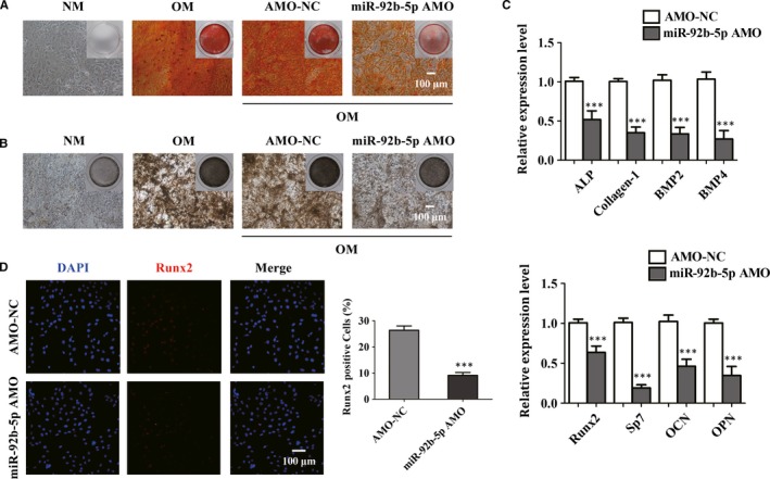
Osteoporosis is closely associated with the dysfunction of bone metabolism, which is caused by the imbalance between new bone formation and bone resorption. Osteogenic differentiation plays a vital role in maintaining the balance of bone microenvironment. The present study investigated whether melatonin participated in the osteogenic commitment of bone marrow mesenchymal stem cells (BMSCs) and further explored its underlying mechanisms. Our data showed that melatonin exhibited the capacity of regulating osteogenic differentiation of BMSCs, which was blocked by its membrane receptor inhibitor luzindole. Further study demonstrated that the expression of miR-92b-5p was up-regulated in BMSCs after administration of melatonin, and transfection of miR-92b-5p accelerated osteogenesis of BMSCs. In contrast, silence of miR-92b-5p inhibited the osteogenesis of BMSCs. The increase in osteoblast differentiation of BMSCs caused by melatonin was attenuated by miR-92b-5p AMO as well. Luciferase reporter assay, real-time qPCR analysis and western blot analysis confirmed that miR-92b-5p was involved in osteogenesis by directly targeting intracellular adhesion molecule-1 (ICAM-1). Melatonin improved the expression of miR-92b-5p, which could regulate the differentiation of BMSCs into osteoblasts by targeting ICAM-1. This study provided novel methods for treating osteoporosis.

Keywords: BMSCs; ICAM-1; MiRNA; melatonin; osteogenic differentiation; osteoporosis.


Conflict of interest statement

The authors indicate no potential conflicts of interest.

Figures

Figure 1
Figure 1
The role of melatonin in the osteogenic differentiation of bone marrow mesenchymal stem cells (BMSCs). (A and B) alizarin red S staining (ARS) (A) and alkaline phosphatase (ALP) (B) staining of BMSCs after culture for 14 d or osteogenic differentiation for 14 d. NM, normal culture medium. OM, osteogenic differentiation‐inducing medium. Scale bar = 100 μm. (C) Real‐time qPCR analysis detected mRNA expression of ALP, Collagen‐1, BMP2, BMP4, Runx2, Sp7, OCN, OPN in BMSCs cultured in NM or OM after 14 d. ALP, Alkaline phosphatase. Collagen‐1, collagen type αI. BMP2, bone morphogenetic protein 2. BMP4, bone morphogenetic protein 4. Runx2, runt‐related transcription factor. Sp7, osterix. OCN, osteocalcin. OPN, osteopontin. (D and E) The osteogenic differentiation of BMSCs exposed to different concentrations of melatonin was observed by ARS staining (D) and ALP (E) staining. Scale bar = 100 μm. (F) The osteoblast‐related genes were differentially expressed in BMSCs treated with melatonin at 1 μmol/L, 10 μmol/L or 100 μmol/L Mel. *P < 0.05, **P < 0.01 and ***P < 0.001 compared with controls
Figure 2
Figure 2
10 μmol/L melatonin promotes the osteoblast differentiation, which was abrogated by luzindole. (A) The calcium deposits of bone marrow mesenchymal stem cells (BMSCs) in the presence of normal culture medium (NM), osteogenic differentiation‐inducing medium (OM), 10 μmol/L Mel or 10 μmol/L Mel+luzindole were detected by ARS staining. Scale bar = 100 μm. Luz, luzindole. (B) ALP staining exhibited different areas and depth of colour of BMSCs in different groups after osteogenic differentiation. Scale bar = 100 μm. (C) Real‐time qPCR analysis measured the mRNA expression of osteogenic‐related genes of BMSCs after the treatment of NM, OM, 10 μmol/L or 10 μmol/L Mel Mel+luzindole. *P < 0.05, **P < 0.01 and ***P < 0.001 compared with NM‐treated group
Figure 3
Figure 3
The effect of miR‐92b‐5p on the osteoblastic differentiation of bone marrow mesenchymal stem cells (BMSCs). (A) The expression of miR‐92b‐5p in BMSCs after treatment with various concentrations of melatonin was measured. (B) Real‐time qPCR technique detected the expression of miR‐92b‐5p between normal culture medium (NM)‐treated BMSCs and OM‐treated BMSCs for 14 d. (C and D) ARS staining (C) and ALP (D) staining investigated the ability of osteogenesis in BMSCs transfected with miR‐92b‐5p mimics. Scale bar = 100 μm. (E) MiR‐92b‐5p mimics elevated the mRNA expression level of osteoblast markers after induction for 14 d. NC, negative control. (F) Immunofluorescence staining visualized the percentage of Runx2‐positive cells in the presence of miR‐92b‐5p mimics or mimics‐NC. Scale bar = 100 μm. **P < 0.01 and ***P < 0.001 compared with mimics‐NC
Figure 4
Figure 4
MiR‐92b‐5p AMO inhibits osteogenesis in bone marrow mesenchymal stem cells (BMSCs). (A and B) ARS (A) staining showed that miR‐92b‐5p AMO decreased the mineralization in BMSCs, which was in accordance with the result of alkaline phosphatase (ALP) staining (B). Scale bar = 100 μm. (C) The expression levels of ALP, Collagen‐1, BMP2, BMP4, Runx2, Sp7, OCN and OPN were determined by real‐time qPCR analysis. (D) Immunofluorescence staining visualized the percentage of Runx2‐positive cells. Scale bar = 100 μm. *P < 0.05, **P < 0.01 and ***P < 0.001 compared with BMSCs transfected with AMO‐NC
Figure 5
Figure 5
MiR‐92b‐5p AMO reversed melatonin‐induced promotion of the differentiation into osteoblasts. (A and B) The increased in osteogenesis by 10 μmol/L Mel was inhibited by miR‐92b‐5p AMO transfection. Scale bar = 100 μm. (C) The expression of osteoblast marker genes was up‐regulated by melatonin treatment, but reversed by the knockdown of AMO. *P < 0.05, **P < 0.01 and ***P < 0.001 compared with NC
Figure 6
Figure 6
MiR‐92b‐5p reduces the osteoporosis‐induced dysfunction of osteogenesis in bone marrow mesenchymal stem cells (BMSCs). (A) Osteogenic ability of BMSCs of OVX‐induced osteoporotic mice was analysed by alizarin red S (ARS) and alkaline phosphatase (ALP) staining. Scale bar = 100 μm. (B) Real‐time qPCR was used to examine the expression of osteoblast‐related genes. (C) The expression of miR‐92b‐5p in bone tissues (left) and BMSCs (right) from Sham and OVX group. (D and E) BMSCs from Sham and OVX group were cultured in osteogenic differentiation‐inducing medium (OM) and subjected to ARS staining (left) and ALP (right) staining. Scale bar = 100 μm. (F) The percentage of Runx2‐positive cells in total BMSCs isolated from Sham and OVX mice was calculated. Scale bar = 100 μm. **P < 0.01 and ***P < 0.001 compared with cells treated with negative control
Figure 7
Figure 7
MiR‐92b‐5p regulates osteogenesis by targeting ICAM‐1. (A) The binding sequence of miR‐92b‐5p and ICAM‐1. (B) The luciferase reporter assay was used to analyse the regulation of miR‐92b‐5p and ICAM‐1. (C) Real‐time qPCR analysis indicated miR‐92b‐5p regulated the expression of ICAM‐1. (D) Western blot was performed to detect the protein expression of ICAM‐1 after transfection of miR‐92b‐5p. ***P < 0.001 compared with cells in the presence of NC

Similar articles

Cited by

References

    1. Ferrari SL. Osteoporosis: romosozumab to rebuild the foundations of bone strength. Nat Rev Rheumatol. 2018;14:128. - PubMed
    1. Tsourdi E, Hofbauer LC. Denosumab: a new treatment option for glucocorticoid‐induced osteoporosis. Lancet Diabetes Endocrinol. 2018;6:428‐429. - PubMed
    1. Lv YJ, Yang Y, Sui BD, et al. Resveratrol counteracts bone loss via mitofilin‐mediated osteogenic improvement of mesenchymal stem cells in senescence‐accelerated mice. Theranostics. 2018;8:2387‐2406. - PMC - PubMed
    1. Frontini‐López YR, Gojanovich AD, Masone D, Bustos DM, Uhart M. Adipose‐derived mesenchymal stem/stromal cells: from the lab bench to the basic concepts for clinical translation. Biocell. 2018;42(3):67‐77.
    1. Balani DH, Ono N, Kronenberg HM. Parathyroid hormone regulates fates of murine osteoblast precursors in vivo. J Clin Investig. 2017;127:3327‐3338. - PMC - PubMed

Publication types

MeSH terms