

Histogram analysis of en face scattering coefficient map predicts malignancy in human ovarian tissue
- PMID: 31304678
- PMCID: PMC7982142
- DOI: 10.1002/jbio.201900115
Histogram analysis of en face scattering coefficient map predicts malignancy in human ovarian tissue
Abstract
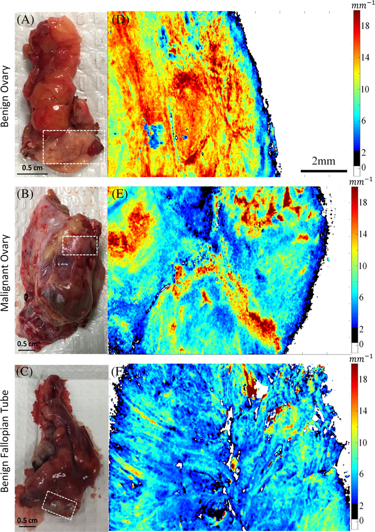
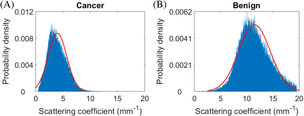
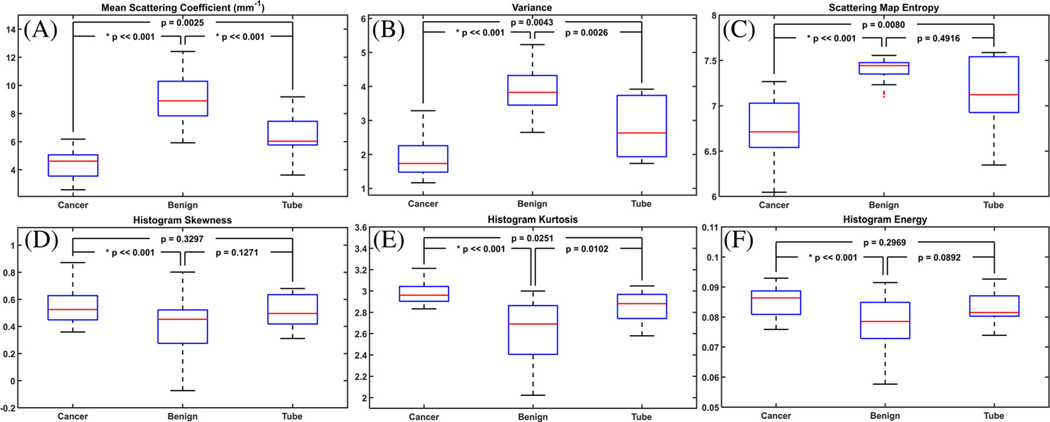
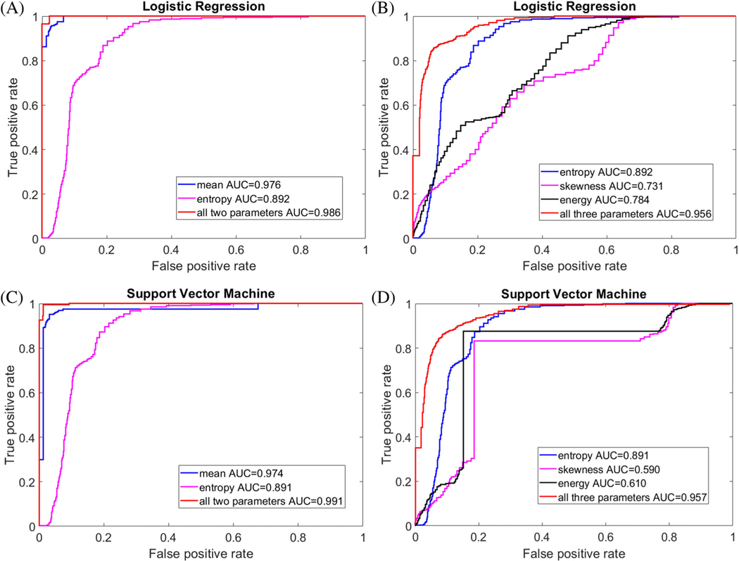
Ovarian cancer is a heterogeneous disease at the molecular and histologic level. Optical coherence tomography (OCT) is able to map ovarian tissue optical properties and heterogeneity, which has been proposed as a feature to aid in diagnosis of ovarian cancer. In this manuscript, depth-resolved en face scattering maps of malignant ovaries, benign ovaries, and benign fallopian tubes obtained from 20 patients are provided to visualize the heterogeneity of ovarian tissues. Six features are extracted from histograms of scattering maps. All features are able to statistically distinguish benign from malignant ovaries. Two prediction models were constructed based on these features: a logistic regression model (LR) and a support vector machine (SVM). The optimal set of features is mean scattering coefficient and scattering map entropy. The LR achieved a sensitivity and specificity of 97.0% and 97.8%, and SVM demonstrated a sensitivity and specificity of 99.6% and 96.4%. Our initial results demonstrate the feasibility of using OCT as an "optical biopsy tool" for detecting the microscopic scattering changes associated with neoplasia in human ovarian tissue.
Keywords: cancer prediction; optical coherence tomography; ovarian cancer; scattering coefficient map.
© 2019 WILEY-VCH Verlag GmbH & Co. KGaA, Weinheim.
Figures
Similar articles
-
Depth-resolved attenuation mapping of the human ovary and fallopian tube using optical coherence tomography.J Biophotonics. 2023 Jun;16(6):e202300002. doi: 10.1002/jbio.202300002. Epub 2023 Mar 21. J Biophotonics. 2023. PMID: 36916760 Free PMC article.
-
Quantitative analysis of angle-resolved scattering properties of ovarian tissue using optical coherence tomography.J Biomed Opt. 2012 Sep;17(9):90503-1. doi: 10.1117/1.JBO.17.9.090503. J Biomed Opt. 2012. PMID: 23085900 Free PMC article.
-
Diagnosing colorectal abnormalities using scattering coefficient maps acquired from optical coherence tomography.J Biophotonics. 2021 Jan;14(1):e202000276. doi: 10.1002/jbio.202000276. Epub 2020 Oct 22. J Biophotonics. 2021. PMID: 33064368 Free PMC article.
-
An overview of optical coherence tomography for ovarian tissue imaging and characterization.Wiley Interdiscip Rev Nanomed Nanobiotechnol. 2015 Jan-Feb;7(1):1-16. doi: 10.1002/wnan.1306. Epub 2014 Oct 20. Wiley Interdiscip Rev Nanomed Nanobiotechnol. 2015. PMID: 25329515 Free PMC article. Review.
-
En-face optical coherence tomography for the detection of cancer in prostatectomy specimens: Quantitative analysis in 20 patients.J Biophotonics. 2020 Jun;13(6):e201960105. doi: 10.1002/jbio.201960105. Epub 2020 Mar 30. J Biophotonics. 2020. PMID: 32049426 Review.
Cited by
-
Enhanced 3D visualization of human fallopian tube morphology using a miniature optical coherence tomography catheter.Biomed Opt Express. 2023 Jun 12;14(7):3225-3233. doi: 10.1364/BOE.489708. eCollection 2023 Jul 1. Biomed Opt Express. 2023. PMID: 37497483 Free PMC article.
-
Optical Resolution Photoacoustic Microscopy of Ovary and Fallopian Tube.Sci Rep. 2019 Oct 4;9(1):14306. doi: 10.1038/s41598-019-50743-7. Sci Rep. 2019. PMID: 31586106 Free PMC article.
-
Multiclass risk models for ovarian malignancy: an illustration of prediction uncertainty due to the choice of algorithm.BMC Med Res Methodol. 2023 Nov 24;23(1):276. doi: 10.1186/s12874-023-02103-3. BMC Med Res Methodol. 2023. PMID: 38001421 Free PMC article.
-
Depth-resolved attenuation mapping of the human ovary and fallopian tube using optical coherence tomography.J Biophotonics. 2023 Jun;16(6):e202300002. doi: 10.1002/jbio.202300002. Epub 2023 Mar 21. J Biophotonics. 2023. PMID: 36916760 Free PMC article.
-
Optical coherence tomography for dynamic investigation of mammalian reproductive processes.Mol Reprod Dev. 2023 Jan;90(1):3-13. doi: 10.1002/mrd.23665. Epub 2022 Dec 27. Mol Reprod Dev. 2023. PMID: 36574640 Free PMC article. Review.
References
-
- Siegel RL, Miller KD, Jemal A, CA Cancer J. Clin. 2019, 69 (1), 7. - PubMed
-
- van Nagell JR Jr, DePriest PD, Reedy MB, Gallion HH, Ueland FR, Pavlik EJ, Kryscio RJ, Gynecol. Oncologia 2000, 77(3), 350. - PubMed
-
- Rebbeck TR, Lynch HT, Neuhausen SL, Narod SA, van’t Veer L, Garber JE, Evans G, Isaacs C, Daly MB, Matloff E, Olopade OI, Weber BL, N. Engl. J. Med. 2002, 346(21), 1616. - PubMed
Publication types
MeSH terms
Grants and funding
LinkOut - more resources
Full Text Sources
Medical
Research Materials
