

Light-Induced Dimerization Approaches to Control Cellular Processes
- PMID: 31304989
- PMCID: PMC6790656
- DOI: 10.1002/chem.201900562
Light-Induced Dimerization Approaches to Control Cellular Processes
Abstract
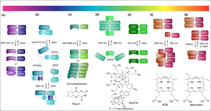
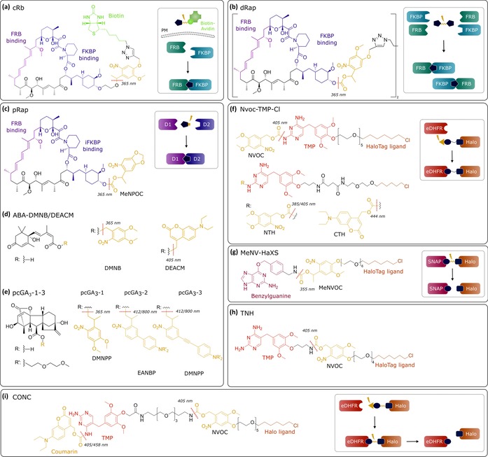
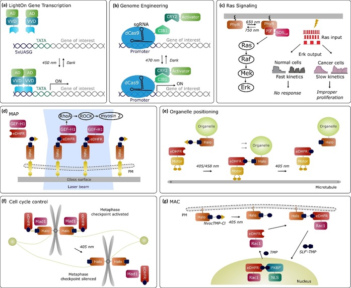
Light-inducible approaches provide a means to control biological systems with spatial and temporal resolution that is unmatched by traditional genetic perturbations. Recent developments of optogenetic and chemo-optogenetic systems for induced proximity in cells facilitate rapid and reversible manipulation of highly dynamic cellular processes and have become valuable tools in diverse biological applications. New expansions of the toolbox facilitate control of signal transduction, genome editing, "painting" patterns of active molecules onto cellular membranes, and light-induced cell cycle control. A combination of light- and chemically induced dimerization approaches have also seen interesting progress. Herein, an overview of optogenetic systems and emerging chemo-optogenetic systems is provided, and recent applications in tackling complex biological problems are discussed.
Keywords: chemo-optogenetics; dimerization; optogenetics; photochemistry; proteins.
© 2019 The Authors. Published by Wiley-VCH Verlag GmbH & Co. KGaA.
Conflict of interest statement
The authors declare no conflict of interest.
Figures
Similar articles
-
Tunable and Photoswitchable Chemically Induced Dimerization for Chemo-optogenetic Control of Protein and Organelle Positioning.Angew Chem Int Ed Engl. 2018 Jun 4;57(23):6796-6799. doi: 10.1002/anie.201800140. Epub 2018 May 8. Angew Chem Int Ed Engl. 2018. PMID: 29637703 Free PMC article.
-
Visible-Light-Switchable Molecular Glues for Reversible Control of Protein Function.Chemistry. 2025 Mar 12;31(15):e202403808. doi: 10.1002/chem.202403808. Epub 2025 Jan 21. Chemistry. 2025. PMID: 39805011
-
Chemo-optogenetic Dimerization Dissects Complex Biological Processes.Small Methods. 2025 Jul;9(7):e2401271. doi: 10.1002/smtd.202401271. Epub 2025 Jan 15. Small Methods. 2025. PMID: 39815164 Review.
-
Modular Photoswitchable Molecular Glues for Chemo-Optogenetic Control of Protein Function in Living Cells.Angew Chem Int Ed Engl. 2025 Feb 10;64(7):e202416456. doi: 10.1002/anie.202416456. Epub 2025 Jan 21. Angew Chem Int Ed Engl. 2025. PMID: 39777946
-
Optochemical Control of Biological Processes in Cells and Animals.Angew Chem Int Ed Engl. 2018 Mar 5;57(11):2768-2798. doi: 10.1002/anie.201700171. Epub 2018 Feb 1. Angew Chem Int Ed Engl. 2018. PMID: 28521066 Free PMC article. Review.
Cited by
-
Light-regulated gene expression in Bacteria: Fundamentals, advances, and perspectives.Front Bioeng Biotechnol. 2022 Oct 14;10:1029403. doi: 10.3389/fbioe.2022.1029403. eCollection 2022. Front Bioeng Biotechnol. 2022. PMID: 36312534 Free PMC article. Review.
-
Light-activated BioID - an optically activated proximity labeling system to study protein-protein interactions.J Cell Sci. 2023 Oct 1;136(19):jcs261430. doi: 10.1242/jcs.261430. Epub 2023 Oct 11. J Cell Sci. 2023. PMID: 37756605 Free PMC article.
-
Allosteric inactivation of an engineered optogenetic GTPase.Proc Natl Acad Sci U S A. 2023 Apr 4;120(14):e2219254120. doi: 10.1073/pnas.2219254120. Epub 2023 Mar 27. Proc Natl Acad Sci U S A. 2023. PMID: 36972433 Free PMC article.
-
A rapid inducible RNA decay system reveals fast mRNA decay in P-bodies.Nat Commun. 2024 Mar 28;15(1):2720. doi: 10.1038/s41467-024-46943-z. Nat Commun. 2024. PMID: 38548718 Free PMC article.
-
How should we regulate products of new breeding techniques? Opinion of surveyed experts in plant biotechnology.Biotechnol Rep (Amst). 2020 May 5;26:e00460. doi: 10.1016/j.btre.2020.e00460. eCollection 2020 Jun. Biotechnol Rep (Amst). 2020. PMID: 32617264 Free PMC article.
References
Publication types
MeSH terms
Substances
Grants and funding
LinkOut - more resources
Full Text Sources
Other Literature Sources
