

Kinematic Evaluation of the GMK Sphere Implant During Gait Activities: A Dynamic Videofluoroscopy Study
- PMID: 31304995
- PMCID: PMC6851890
- DOI: 10.1002/jor.24416
Kinematic Evaluation of the GMK Sphere Implant During Gait Activities: A Dynamic Videofluoroscopy Study
Erratum in
-
Corrigendum.J Orthop Res. 2020 Sep;38(9):2083. doi: 10.1002/jor.24703. Epub 2020 May 6. J Orthop Res. 2020. PMID: 32779802 Free PMC article. No abstract available.
Abstract
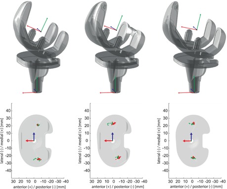
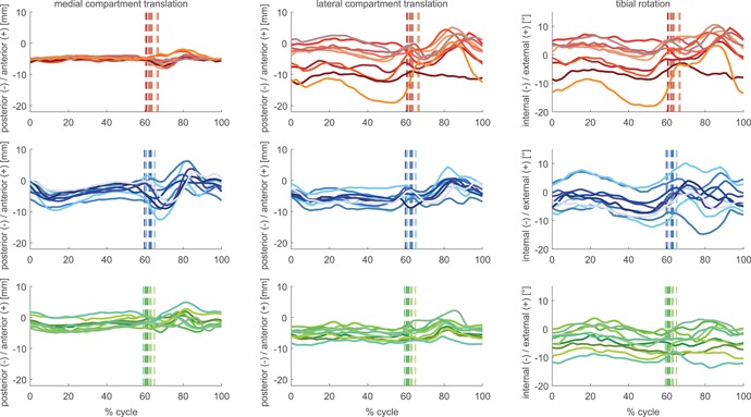
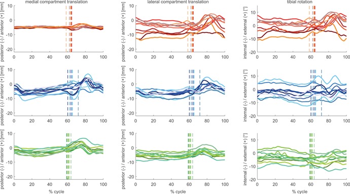
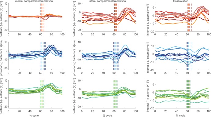
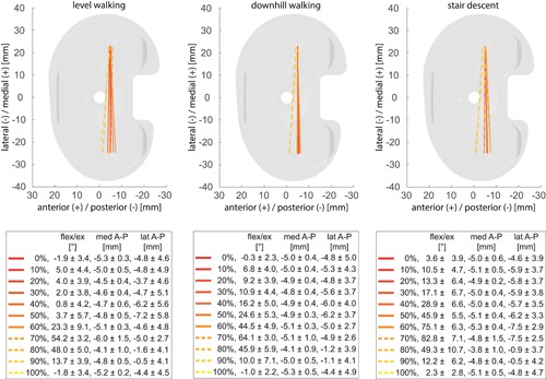
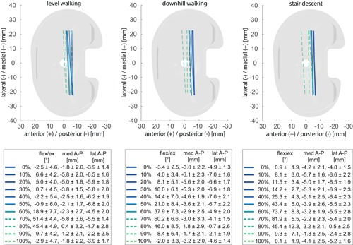
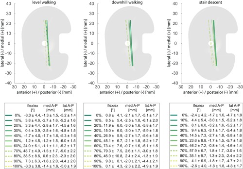
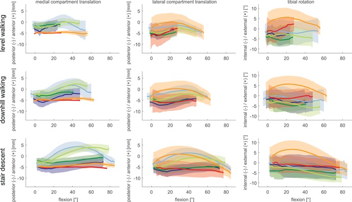
Joint stability is a primary concern in total knee joint replacement. The GMK Sphere prosthesis was specifically designed to provide medial compartment anterior-posterior (A-P) stability, while permitting rotational freedom of the joint through a flat lateral tibial surface. The objective of this study was to establish the changes in joint kinematics introduced by the GMK Sphere prosthesis during gait activities in comparison to conventional posterior-stabilized (PS) fixed-bearing and ultra-congruent (UC) mobile-bearing geometries. The A-P translation and internal/external rotation of three cohorts, each with 10 good outcome subjects (2.9 ± 1.6 years postop), with a GMK Sphere, GMK PS or GMK UC implant were analysed throughout complete cycles of gait activities using dynamic videofluoroscopy. The GMK Sphere showed the smallest range of medial compartment A-P translation for level walking, downhill walking, and stair descent (3.6 ± 0.9 mm, 3.1 ± 0.8 mm, 3.9 ± 1.3 mm), followed by the GMK UC (5.7 ± 1.0 mm, 8.0 ± 1.7 mm, 8.7 ± 1.9 mm) and the GMK PS (10.3 ± 2.2 mm, 10.1 ± 2.6 mm, 11.6 ± 1.6 mm) geometries. The GMK Sphere exhibited the largest range of lateral compartment A-P translation (12.1 ± 2.2 mm), and the largest range of tibial internal/external rotation (13.2 ± 2.2°), both during stair descent. This study has shown that the GMK Sphere clearly restricts A-P motion of the medial condyle during gait activities while still allowing a large range of axial rotation. The additional comparison against the conventional GMK PS and UC geometries, not only demonstrates that implant geometry is a key factor in governing tibio-femoral kinematics, but also that the geometry itself probably plays a more dominant role for joint movement than the type of gait activity. © 2019 The Authors. Journal of Orthopaedic Research® published by Wiley Periodicals, Inc. on behalf of Orthopaedic Research Society. J Orthop Res 37:2337-2347, 2019.
Keywords: gait activities; medial congruent; moving fluoroscope; single plane fluoroscopy; total knee arthroplasty.
© 2019 The Authors. Journal of Orthopaedic Research® published by Wiley Periodicals, Inc. on behalf of Orthopaedic Research Society.
Figures
Similar articles
-
ISB clinical biomechanics award winner 2021: Tibio-femoral kinematics of natural versus replaced knees - A comparison using dynamic videofluoroscopy.Clin Biomech (Bristol). 2022 Jun;96:105667. doi: 10.1016/j.clinbiomech.2022.105667. Epub 2022 May 20. Clin Biomech (Bristol). 2022. PMID: 35636308
-
Can a total knee arthroplasty be both rotationally unconstrained and anteroposteriorly stabilised? A pulsed fluoroscopic investigation.Bone Joint Res. 2016 Mar;5(3):80-6. doi: 10.1302/2046-3758.53.2000621. Bone Joint Res. 2016. PMID: 26965166 Free PMC article.
-
Tibio-femoral kinematics of the healthy knee joint throughout complete cycles of gait activities.J Biomech. 2020 Sep 18;110:109915. doi: 10.1016/j.jbiomech.2020.109915. Epub 2020 Jul 18. J Biomech. 2020. PMID: 32827791
-
Posterior-stabilized inserts are preferable to cruciate-substituting ultracongruent inserts due to more favourable kinematics and stability.Knee Surg Sports Traumatol Arthrosc. 2018 Nov;26(11):3300-3310. doi: 10.1007/s00167-018-4872-z. Epub 2018 Feb 19. Knee Surg Sports Traumatol Arthrosc. 2018. PMID: 29459998
-
A novel approach to knee kinematics.Am J Orthop (Belle Mead NJ). 2001 Apr;30(4):287-93. Am J Orthop (Belle Mead NJ). 2001. PMID: 11334450 Review.
Cited by
-
Effect of the soft tissue artifact on marker measurements and on the calculation of the helical axis of the knee during a squat movement: A study on the CAMS-Knee dataset.Med Eng Phys. 2022 Dec;110:103915. doi: 10.1016/j.medengphy.2022.103915. Epub 2022 Oct 29. Med Eng Phys. 2022. PMID: 36564140 Free PMC article.
-
Kinematically Aligned Total Knee Arthroplasty with Patient-Specific Instrument.Yonsei Med J. 2020 Mar;61(3):201-209. doi: 10.3349/ymj.2020.61.3.201. Yonsei Med J. 2020. PMID: 32102120 Free PMC article. Review.
-
More passive internal tibial rotation with posterior cruciate ligament retention than with excision in a medial pivot TKA implanted with unrestricted caliper verified kinematic alignment.Knee Surg Sports Traumatol Arthrosc. 2023 Mar;31(3):852-860. doi: 10.1007/s00167-021-06840-0. Epub 2021 Dec 18. Knee Surg Sports Traumatol Arthrosc. 2023. PMID: 34921630 Free PMC article.
-
An insert with less than spherical medial conformity causes a loss of passive internal rotation after calipered kinematically aligned TKA.Arch Orthop Trauma Surg. 2021 Dec;141(12):2287-2294. doi: 10.1007/s00402-021-04054-0. Epub 2021 Jul 15. Arch Orthop Trauma Surg. 2021. PMID: 34264381 Free PMC article.
-
Conceptual foundations of a REFRAME-based approach to discriminate across total knee implant designs based on the positions of functional centres of rotation.Sci Rep. 2025 Jan 4;15(1):834. doi: 10.1038/s41598-024-84522-w. Sci Rep. 2025. PMID: 39755774 Free PMC article.
References
-
- Dennis DA, Komistek RD, Mahfouz MR. 2003. In vivo fluoroscopic analysis of fixed‐bearing total knee replacements. Clin Orthop Relat Res 410:114–130. - PubMed
-
- Blaha JD. 2004. The rationale for a total knee implant that confers anteroposterior stability throughout range of motion. J Arthroplasty 19:22–26. - PubMed
-
- Pritchett JW. 2011. Patients prefer a bicruciate‐retaining or the medial pivot total knee prosthesis. J Arthroplasty 26:224–228. - PubMed
-
- Iwaki H, Pinskerova V, Freeman MA. 2000. Tibiofemoral movement 1: the shapes and relative movements of the femur and tibia in the unloaded cadaver knee. J Bone Joint Surg Br 82:1189–1195. - PubMed
Publication types
MeSH terms
Grants and funding
LinkOut - more resources
Full Text Sources
