Case Reports
. 2019 Oct 7;152(5):590-607.
doi: 10.1093/ajcp/aqz080.

Migration of Talc From the Perineum to Multiple Pelvic Organ Sites

Affiliations
Case Reports

Migration of Talc From the Perineum to Multiple Pelvic Organ Sites

Sandra A McDonald et al. Am J Clin Pathol. .

Abstract

Objectives: Genital talc use is associated with increased risk for ovarian carcinoma in epidemiologic studies. Finding talc in pelvic tissues in women with ovarian carcinoma who have used talc is important in documenting exposure and assessing talc's biologic potential, but tissue-based morphology studies have been rarely reported.

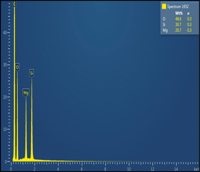
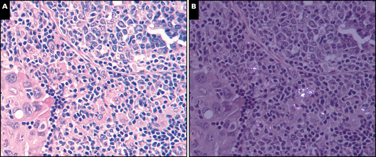
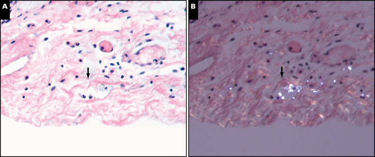
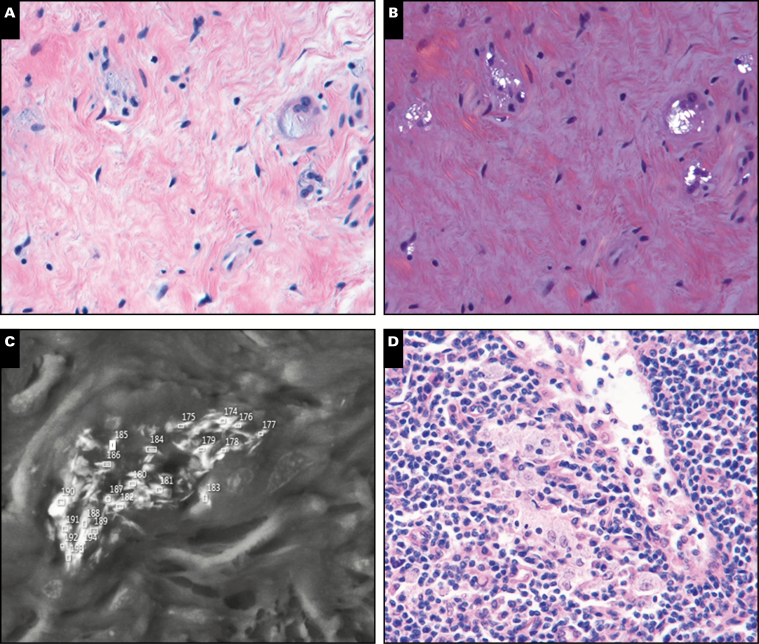
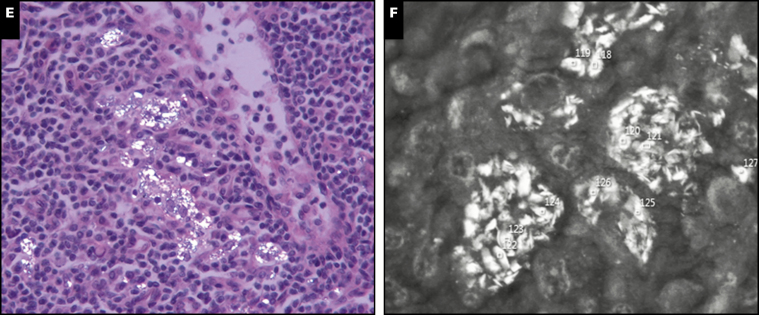
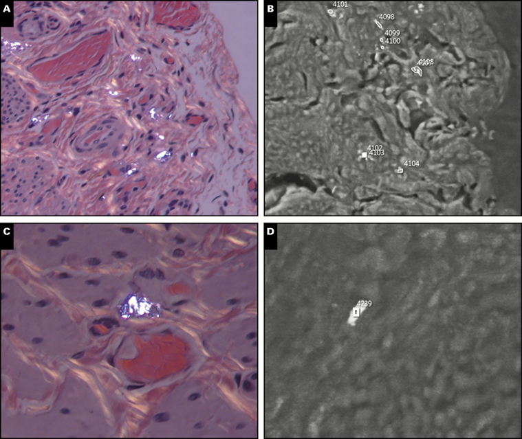
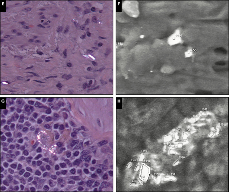
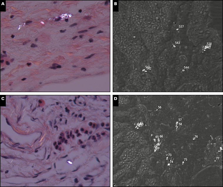
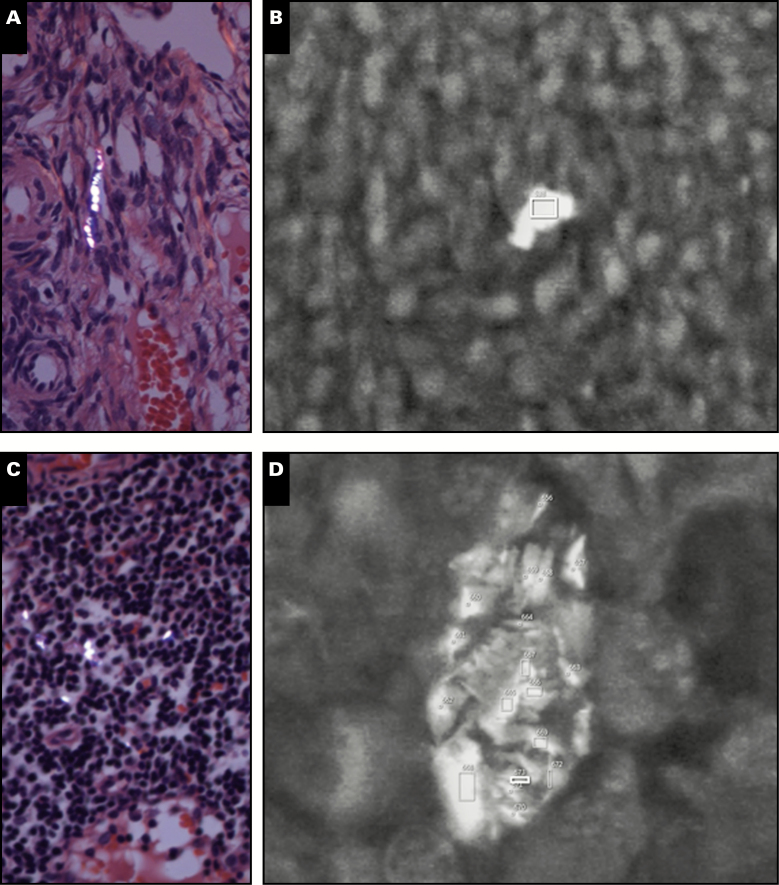
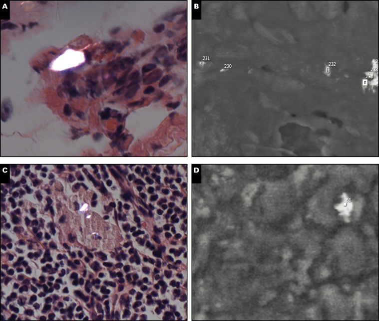
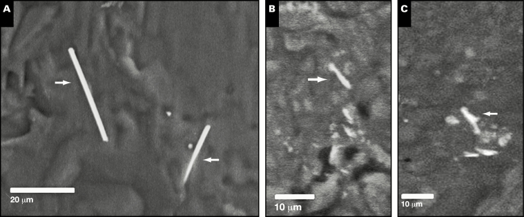
Methods: We report five patient cases with documented perineal talc use, each of whom had talc (by both polarized light and scanning electron microscopy) in multiple pelvic sites distant from the perineum. Six negative-exposure control patients were also analyzed.

Results: Talc particles were found in exposed patients, typically within two or more of the following locations: pelvic region lymph nodes, cervix, uterine corpus, fallopian tubes, and ovaries.

Conclusions: Our report adds new insights into the biologic potential of talc and suggests additional anatomic sites that should be closely examined for talc by oncologic surgical pathologists in the setting of perineal talc use.

Keywords: Ovarian carcinoma; Pelvic lymph node; Polarized light microscopy; Scanning electron microscopy; Talc.


Figures

Image 1
Image 1
Representative spectrum of talc, showing characteristic magnesium (Mg) and silicon (Si) peaks. The characteristic Mg-Si atomic ratio is 0.75 and atomic weight percent ratio is 0.649, and particles are considered to be talc if their Mg-Si ratio falls within 5% of this theoretical value (0.649).
Image 2
Image 2
Regular light microscopy (A) and polarized light microscopy (B) of left pelvic lymph node in patient 2, same field of view, showing juxtaposition of birefringent particles in macrophages, metastatic carcinoma, and uninvolved lymph node parenchyma. This particular area was not analyzed by scanning electron microscopy, but based on the findings in other histologic regions, much of this birefringent material is likely talc. (H&E, ×400)
Image 3
Image 3
Regular light microscopy (A) and polarized light microscopy (B) of the uterine serosa in patient 3, same field of view, showing serosal fibrovascular tissue and abundant birefringent particles that are seen in one lymphvascular space (arrows). This serosal birefringent material was shown to be talc by scanning electron microscopy/energy-dispersive X-ray analysis (see Image 6). (H&E, ×200)
Image 4
Image 4
Representative photomicrographs for patient 1. A, Deep exocervical soft tissue with collections of macrophages in dense collagenous tissue. A few macrophages were multinucleate and showed slightly glassy grayish cytoplasm (H&E, ×400). B, Same histologic field as A, under polarized light microscopy, showing collections of macrophages with numerous birefringent cytoplasmic particles 1 to 10 μm in diameter (H&E, ×400). C, Scanning electron microscopy (SEM, ×500) with backscattered electron imaging from the same general area as in A and B but a different histologic section, showing numerous backscattered electron-positive particulates within the cytoplasm of macrophages, similar to A, the majority of which had a spectrum characteristic of talc. D, Right pelvic lymph node with aggregates of intranodal macrophages (H&E, ×400). E, Same histologic field as D, under polarized light microscopy, with numerous birefringent particles similar in size and appearance to those seen in the exocervix, within intranodal macrophages (H&E, ×400). F, SEM (×500) from the same general area as D and E but in a different histologic section, showing numerous backscattered electron-positive particulates within the cytoplasm of macrophages, similar to C, and the majority having a spectrum characteristic of talc.
Image 4
Image 4
Representative photomicrographs for patient 1. A, Deep exocervical soft tissue with collections of macrophages in dense collagenous tissue. A few macrophages were multinucleate and showed slightly glassy grayish cytoplasm (H&E, ×400). B, Same histologic field as A, under polarized light microscopy, showing collections of macrophages with numerous birefringent cytoplasmic particles 1 to 10 μm in diameter (H&E, ×400). C, Scanning electron microscopy (SEM, ×500) with backscattered electron imaging from the same general area as in A and B but a different histologic section, showing numerous backscattered electron-positive particulates within the cytoplasm of macrophages, similar to A, the majority of which had a spectrum characteristic of talc. D, Right pelvic lymph node with aggregates of intranodal macrophages (H&E, ×400). E, Same histologic field as D, under polarized light microscopy, with numerous birefringent particles similar in size and appearance to those seen in the exocervix, within intranodal macrophages (H&E, ×400). F, SEM (×500) from the same general area as D and E but in a different histologic section, showing numerous backscattered electron-positive particulates within the cytoplasm of macrophages, similar to C, and the majority having a spectrum characteristic of talc.
Image 5
Image 5
Representative photomicrographs for patient 2. A, Uterine serosa showing numerous birefringent particles 1 to 10 mm in diameter within soft tissue and macrophages (H&E, ×200). B, Scanning electron microscopy (SEM) corresponding to A, showing numerous backscattered electron-positive particles (×500), the majority with an energy-dispersive X-ray analysis (EDX) spectrum characteristic of talc. C, Fibromuscular soft tissue near fallopian tube, showing a macrophage with abundant intracellular birefringent material similar to that seen in A (H&E, ×400). D, SEM of the same region as C, showing a backscattered electron-positive particle approximately 5 μm in diameter that proved to be talc using EDX (×500). E, Birefringent particle seen in soft tissue of ovary, with mixed inflammation and fibrosis in the general background (H&E, ×400). F, SEM showing several backscattered electron-positive particles within the same region as E (ovary). The particles were irregularly shaped, less than 7 μm in diameter, and on EDX analysis showed the characteristic spectrum of talc (Image 1) (×500). G, Left pelvic lymph node with numerous birefringent particles similar in size and appearance to those seen in the uterine serosa, within intranodal macrophages (H&E, ×400). H, SEM from the same general area as E but in a different histologic section, showing numerous backscattered electron-positive particulates within the cytoplasm of macrophages, similar to E, with most having an EDX spectrum characteristic of talc (×500).
Image 5
Image 5
Representative photomicrographs for patient 2. A, Uterine serosa showing numerous birefringent particles 1 to 10 mm in diameter within soft tissue and macrophages (H&E, ×200). B, Scanning electron microscopy (SEM) corresponding to A, showing numerous backscattered electron-positive particles (×500), the majority with an energy-dispersive X-ray analysis (EDX) spectrum characteristic of talc. C, Fibromuscular soft tissue near fallopian tube, showing a macrophage with abundant intracellular birefringent material similar to that seen in A (H&E, ×400). D, SEM of the same region as C, showing a backscattered electron-positive particle approximately 5 μm in diameter that proved to be talc using EDX (×500). E, Birefringent particle seen in soft tissue of ovary, with mixed inflammation and fibrosis in the general background (H&E, ×400). F, SEM showing several backscattered electron-positive particles within the same region as E (ovary). The particles were irregularly shaped, less than 7 μm in diameter, and on EDX analysis showed the characteristic spectrum of talc (Image 1) (×500). G, Left pelvic lymph node with numerous birefringent particles similar in size and appearance to those seen in the uterine serosa, within intranodal macrophages (H&E, ×400). H, SEM from the same general area as E but in a different histologic section, showing numerous backscattered electron-positive particulates within the cytoplasm of macrophages, similar to E, with most having an EDX spectrum characteristic of talc (×500).
Image 6
Image 6
Representative photomicrographs for patient 3. A, Uterine serosa showing numerous birefringent particles 1 to 10 μm in diameter within soft tissue and macrophages (H&E, ×400). B, Scanning electron microscopy (SEM) corresponding to A, showing numerous backscattered electron-positive particles (×500), most with an energy-dispersive X-ray analysis spectrum of talc similar to Image 1. C, Ovarian stroma showing a birefringent particle within soft tissue but close to blood vessel. Other birefringent particles were seen in different microscopic fields in this section. D, SEM showing several backscattered electron-positive particles within the same tissue (but different section) corresponding to C. These backscattered electron-positive particles showed a spectrum characteristic of talc (Image 1) (×500).
Image 7
Image 7
Representative photomicrograph for patient 4. A, Tissue of the right ovary with a string-like arrangement of multiple birefringent particles (1-5 μm in greatest dimension) within ovarian stromal tissue (H&E, ×400). B, Scanning electron microscopy (SEM) showing an irregularly shaped particle, which in backscatter mode is about 6 μm in diameter. Energy-dispersive X-ray analysis of this particle showed the typical spectrum of talc (Image 1) (×1,000). C, Right pelvic lymph node tissue with approximately eight birefringent particles (each ~2 μm in greatest dimension or less) visible in the same plane of focus with the cells of the lymph node. Many of these particles are clearly within macrophage cells in the lymph node. D, SEM of the same right pelvic lymph node tissue (but a different section) showing numerous backscattered electron-positive particles within the cytoplasm of a macrophage, similar to the light microscopic morphology in C. These particles had the characteristic spectrum of talc (Image 1) (×1,000).
Image 8
Image 8
Representative photomicrograph for patient 5. A, Birefringent particle approximately 7 μm in diameter, found in the soft tissues around the fallopian tube, and associated with chronic inflammation (H&E, ×400). B, Scanning electron microscopy (SEM) showing numerous backscattered electron-positive particles within the soft tissues around the same fallopian tube tissue as in A but a different tissue section (×500). C, Two birefringent particles within a left pelvic lymph node (H&E, ×400). D, SEM of the same pelvic lymph node tissue as in C, showing an irregularly shaped, backscattered electron-positive particle less than 5 μm in diameter, which showed the characteristic spectrum of talc.
Image 9
Image 9
Representative fibers and fiber-like particles (≥5:1 aspect ratio) found in our patient analysis (all photos are scanning electron microscopy with backscattered electron imaging, ×500). A, Long aspect ratio fibers (arrows) with a chemical signature of calcium (Ca), magnesium (Mg), aluminum (Al), iron, and silicon (Si) found in patient case 2 (right ovary). Fiber at right is seen to be extending into tissue where it disappears from view; thus, its aspect ratio may be higher than what is visible. For the total of four long aspect ratio fibers (10:1 or greater) that we found in our study, based on atomic weight percent calculations, average Mg/Si was 0.241 and Ca/Si was 1.03, where the respective ratios expected for tremolite are 0.542 and 0.357. Average Al/Si for the four fibers was 0.327, whereas no Al is expected for tremolite. B, Talc fiber-like particle (arrow) with an approximately 6:1 aspect ratio from case 2 (uterus). C, Talc fiber-like particle (arrow) from case 3 (uterus), with an approximately 6:1 aspect ratio. Particles in B and C had Mg-Si spectra with atomic weight percent ratios within 5% of the theoretical value of 0.649 for talc, similar to Image 1. In C, the other nearby backscattered electron-positive particles were also talc but did not meet the 5:1 aspect ratio threshold for a fibrous morphology.

Similar articles

Cited by

References

    1. Cramer DW, Welch WR, Scully RE, et al. . Ovarian cancer and talc: a case-control study. Cancer. 1982;50:372-376. - PubMed
    1. Cramer DW, Liberman RF, Titus-Ernstoff L, et al. . Genital talc exposure and risk of ovarian cancer. Int J Cancer. 1999;81:351-356. - PubMed
    1. Cramer DW, Vitonis AF, Terry KL, et al. . The association between talc use and ovarian cancer: a retrospective case-control study in two US states. Epidemiology. 2016;27:334-346. - PMC - PubMed
    1. Schildkraut JM, Abbott SE, Alberg AJ, et al. . Association between body powder use and ovarian cancer: the African American Cancer Epidemiology Study (AACES). Cancer Epidemiol Biomarkers Prev. 2016;25:1411-1417. - PMC - PubMed
    1. Terry KL, Karageorgi S, Shvetsov YB, et al. ; Australian Cancer Study (Ovarian Cancer); Australian Ovarian Cancer Study Group; Ovarian Cancer Association Consortium Genital powder use and risk of ovarian cancer: a pooled analysis of 8525 cases and 9859 controls. Cancer Prev Res (Phila). 2013;6:811-821. - PMC - PubMed

Publication types

MeSH terms