. 2019;70(4):1025-1040.
doi: 10.3233/JAD-190161.

Effects of Amylin Against Amyloid-β-Induced Tauopathy and Synapse Loss in Primary Neurons

Affiliations

Effects of Amylin Against Amyloid-β-Induced Tauopathy and Synapse Loss in Primary Neurons

Qini Gan et al. J Alzheimers Dis. 2019.

Abstract

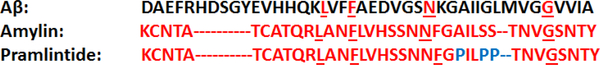
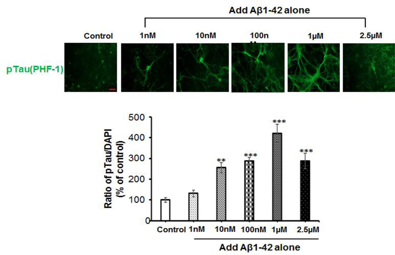
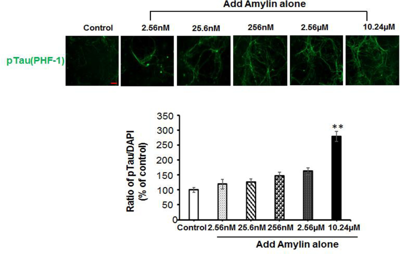
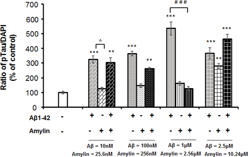
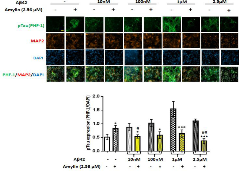
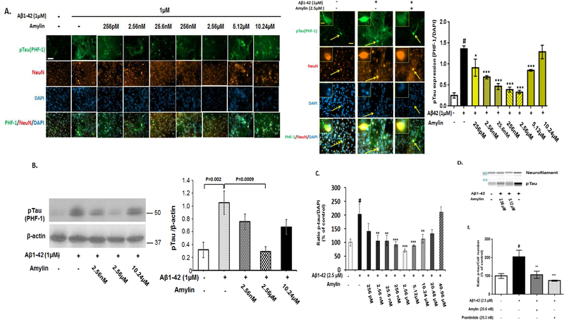
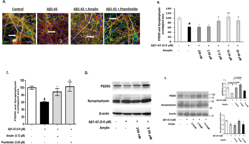
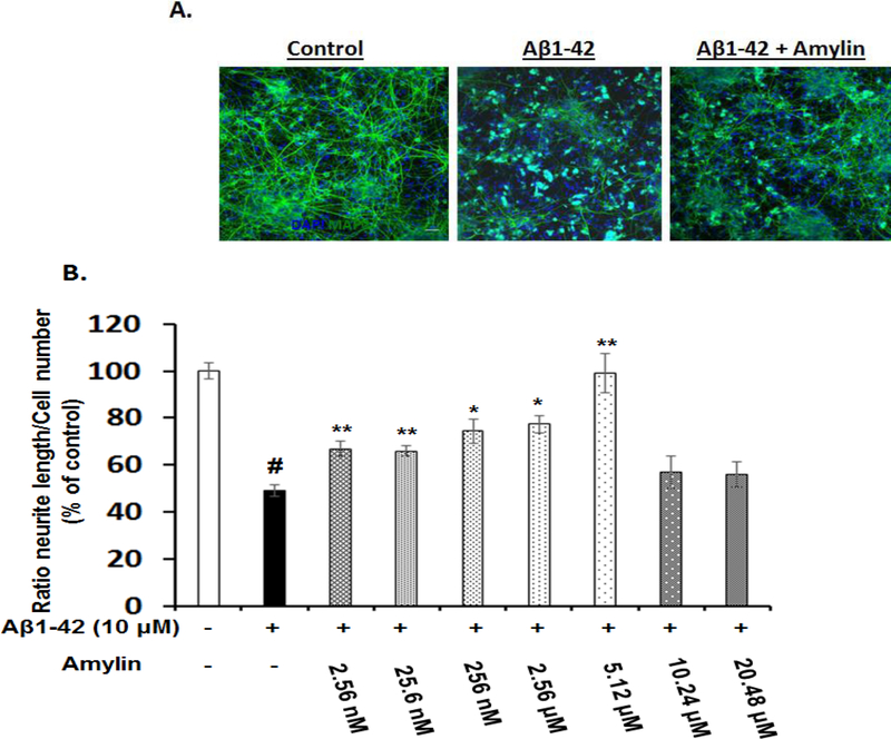
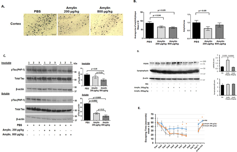
Recent studies demonstrate that peripheral amylin treatment reduces pathology in mouse models of Alzheimer's disease (AD). However, soluble and aggregated amylin are distinct species; while amylin is a physiological neuropeptide, amylin aggregation is a pathological factor for diabetes. We thus hypothesized that because of their similarity in secondary structures, amylin antagonizes amyloid-β peptide (Aβ)-induced AD pathology in neurons with a dose-dependent pattern. To test the hypothesis, we conducted both in vitro and in vivo experiments with different doses of amylin and with its analog, pramlintide. Here we report that a high concentration of either Aβ or amylin alone induced tau phosphorylation (pTau) in primary neurons. Interestingly, with a low concentration, amylin had direct effects to reverse the Aβ-induced pTau, as well as damaged neuronal synapses and neurite disorganization. However, when the concentration was high (10.24 μM), amylin lost the effects against the Aβ-induced cellular AD pathology and, together with Aβ, worsened tauopathy in neurons. In the 5XFAD AD mouse model, daily peripheral amylin treatment with a low dose (200 μg/kg) more effectively reduced amyloid burden, and increased synapse, but with a high dose (800 μg/kg), it more effectively reduced tauopathy. Correspondingly, amylin treatment improved learning and memory in these mice. It demonstrates that amylin has a dose-dependent U-shape effect against AD pathogenesis. Within a physiological range, amylin is a neuroprotective hormone against AD in neurons; but when both Aβ and amylin concentrations are elevated, imbalance of Aβ and amylin may contribute to brain AD pathogenesis.

Keywords: Alzheimer’s disease; U-shape; amylin; amyloid-β peptide; pramlintide; synapse; tauopathy.


Figures

Figure 1.
Figure 1.. Primary amino acid sequences of Aβ and amylin-type peptides
The amino acid sequences of Aβ1–42, amylin and pramlintide are presented. Amino acids of amylin are in red, and the amino acids of other peptides are in red if they are identical to that in amylin. The different amino acids of pramlintide from amylin are in blue, while the different amino acids of Aβ1–42 from amylin are in black.
Figure 2.
Figure 2.. Either Aβ or amylin alone could induce cellular tau phosphorylation in primary neurons
Mouse cortical neurons were grown and treated with Aβ1–42 alone (A), or human amylin alone (B), or Aβ1–42 plus amylin (C). The neurons were fixed, incubated with the pTau antibody, PHF-1. When the concentration of either peptide was increased, both Aβ1–42 (A) and human amylin (B) alone induced cellular tau phosphorylation (pTau) (Scale bars: 50 μm). The maximum amount of 2.5 μM Aβ1–42 induced less pTau than 1 μM Aβ1–42 due to some neuronal death. The level of tau phosphorylation was normalized by DAPI to allow comparisons among different experimental groups. Values are expressed relative to the controls (untreated mouse primary neurons), which were set as 100%. Values are the mean ± SE from all experiments. For A and B, compared to the control condition, differences with statistical significance are shown with * p<0.05; ** p<0.01; *** p<0.001. For C, compared to the condition of Aβ1–42 alone, the differences of condition of amylin plus Aβ1–42 are shown with statistical significance # p<0.05; ## p<0.01; the differences between the conditions with amylin alone and amylin plus Aβ1–42 are shown with statistical significance ^ p<0.05; ^^ p<0.01. Each experiment was repeated three times and the representative imagings are shown.
Figure 3.
Figure 3.. Preincubation with amylin dramatically reduced Aβ induced cellular tau phosphorylation in primary neurons regardless of increasing amounts of Aβ
Mouse cortical neurons were grown and pretreated without and with amylin (2.56 μM) for 1 hr before adding increasing amounts of aggregated Aβ1–42 for 16 hrs. The neurons were fixed, and incubated with the pTau antibody, PHF-1, to detect cellular tauopathy. The neurons were also immunostained by MAP2 antibody and DAPI (Scale bars: 50 μm). The differences between the absence and the presence of amylin for each concentration of Aβ1–42 are shown with statistical significance * p<0.05; ** p<0.01; *** p<0.001. The differences between the condition with amylin alone and amylin plus Aβ1–42 are shown with statistical significance #p<0.05; ##p<0.01. The experiment was repeated three times and the representative imagings are shown.
Figure 4.
Figure 4.. Amylin’s protective effects against the pathological process of tauopathy in cortical neurons are a dose-dependent, U shaped pattern
Mouse cortical neurons were grown and pretreated without and with increasing concentrations of amylin for 1 hr before adding the fixed amount of aggregated Aβ1–42 (1 μM) for 16 hrs. The neurons were fixed, incubated with the pTau antibody, PHF-1, to detect cellular tauopathy and quantitated. The neurons were immunostained by NeuN antibody and DAPI as well (Scale bars: 50 μm). An indicated (arrow) PHF-1 positive-neuron is showed in a higher-magnification inset in the right panel (Scale bars: 10 μm) (A). Western blots were conducted to reveal the expressions of pTau (PHF-1 antibody) in neurons in the conditions of control, Aβ alone, Aβ plus amylin (B). To confirm the result, we also used rat fetal cortical neurons and conducted the immunohistochemistry (C) as described for mouse cortical neurons except adding aggregated Aβ1–42 (2.5 μM-best concentration to induce tauopathy in rat neurons). Western blot (D) was conducted to confirm the immunohistochemistry in (C). The same concentrations of amylin and pramlintide were added to the rat neurons for 1 hr before adding aggregated Aβ1–42 in the immunostainning experiment for pTau expression (E). The neurons were fixed, incubated with different antibodies to reveal the pathological component of pTau, and quantified, and the representative photographs are shown. # represents the comparisons between the control condition of treatment (PBS) and the condition with adding aggregated Aβ (p<0.001). Compared to the condition with aggregated Aβ, differences in the preincuation of either amylin or pramlintide are shown with statistical significance * p<0.05; ** p<0.01; *** p<0.001. Each experiment was repeated three times and the representative images and the WB gels are shown.
Figure 5.
Figure 5.. Amylin reversed Aβ induced synapse loss in primary neurons
Rat fetal hippocampal neurons were grown and treated with aggregated Aβ1–42 (0.6 μM), causing loss of synapses, defined by co-localization of PSD95 and Synaptophysin, in these neurons. The cultures were treated with control medium or medium containing different concentrations of amylin or pramlintide before adding Aβ1–42. The neurons were then fixed, incubated with different antibodies to reveal PSD95 and Synaptophysin, and quantified. The experiment was repeated three times and the representative images are shown. The photos of controls, Aβ, Aβ+amylin (10.24 μM) and Aβ+pramlintide (5.06 μM) are shown (A) and quantified (C); Aβ+different concentrations of amylin were quantified (B). Western blots were conducted to reveal the expressions of PSD95 and Synaptophysin in primary rat hippocampal neurons. The experiment was repeated three times and the representative WB gels are shown (D). In addition, Western blots of mouse cortical neurons in the conditions of control, Aβ alone, Aβ plus amylin were conducted and quantitated to detect the expressions of PSD95 and Synaptophysin (E). The experiment was repeated four times and the representative WB gels are shown. # represents the condition of synapse loss after adding aggregated Aβ and compared with that in the control condition (p<0.001). Compared to the incubation condition with aggregated Aβ alone, differences with statistical significance are shown with * p<0.05; ** p<0.01.
Figure 6.
Figure 6.. Amylin type peptides’ effects on Aβ induced neurite damage in primary cortical neurons
Rat fetal cortical neurons were grown and treated with aggregated Aβ1–42, causing reduced neurite outgrowth in these neurons. The neurons were fixed, incubated with the antibodies to reveal MAP2 and nuclei, and quantified for the neurite length. The representative photos of controls, Aβ, and Aβ plus amylin (2.56 μM) are shown (A). The average length of neurites in rat neurons which pretreated without and with increasing concentrations of amylin for 1 hour before adding the fixed amount of aggregated Aβ1–42 (10 μM) for 24 hours were quantified (B). # represents the condition of incubation after adding aggregated Aβ and compared with that in the control condition (p<0.001). Compared to the incubation condition with aggregated Aβ alone, differences with statistical significance are shown with * p<0.05; ** p<0.01. The experiment was repeated three times and the representative images are shown.
Figure 7.
Figure 7.. Peripheral amylin treatment with a low vs. a high dose reduces AD pathology and improves learning and memory in an AD mouse model
Amylin treatment of 5XFAD mice reduces the AD pathology and improves their learning and memory. At 4 months of age, 5XFAD mice were treated by i.p. injection of PBS or amylin (200 μg/kg vs. 800 μg/kg) daily for 6 weeks (n = 14–16 per group). (A) Dense-cored Aβ plaque burden is significantly reduced by the amylin 200 μg/kg treatment in the cortex and less so by the amylin 800 μg/kg treatment. The photos of cerebral cortex are shown (Scale bars: 200 μm). (B) Amylin treated (200 μg/kg vs. 800 μg/kg) mice had a significant reduction in Aβ amyloid burden in cortex quantitated by average plaque size, total amyloid area. Again, the dose of 800 μg/kg was less effective than 200 μg/kg. (C) Protein extracts from the brains were fractionated into soluble fraction by TBS extraction and insoluble fraction by the sarkosyl-extraction followed by Western blots with the PFH-1 antibody against pTau and quantitated. Total tau and actin were detected by Western blots and are shown. The amylin treatment with 800 μg/kg was more effective to reduce tauopathy in the brain than the dose of 200 μg/kg. (D) Total protein from the brains was extracted followed by Western blots with the antibody against PSD95 and the one against Synaptophisin. While the amylin treatment with the dose of 200 μg/kg, but not the dose of 800 μg/kg, effectively increased the expression of PSD95, both amylin doses did not influence the expression of Synaptophysin. (E) The amylin treated 5XFAD mice illustrated improved cognition compared to the PBS treatment by showing shortened times in Morris water maze test in finding the hidden platform at day 7 and 8 (D7–8), in memory at day 11 (D11) after the completion of training and skipping two days, and in the probe trial. Mean ± SE was used with *p < 0.05.

Similar articles

Cited by

References

    1. Nishi M, Sanke T, Seino S, Eddy RL, Fan YS, Byers MG, Shows TB, Bell GI, Steiner DF (1989) Human islet amyloid polypeptide gene: complete nucleotide sequence, chromosomal localization, and evolutionary history. Molecular endocrinology (Baltimore, Md.) 3, 1775–1781. - PubMed
    1. Lutz TA (2013) The interaction of amylin with other hormones in the control of eating. Diabetes, obesity & metabolism 15, 99–111. - PubMed
    1. Mietlicki-Baase EG (2016) Amylin-mediated control of glycemia, energy balance, and cognition. Physiol Behav. - PMC - PubMed
    1. Qiu WQ, Zhu H (2014) Amylin and its analogs: a friend or foe for the treatment of Alzheimer’s disease? Frontiers in aging neuroscience 6, 186. - PMC - PubMed
    1. Adler BL, Yarchoan M, Hwang HM, Louneva N, Blair JA, Palm R, Smith MA, Lee HG, Arnold SE, Casadesus G (2014) Neuroprotective effects of the amylin analogue pramlintide on Alzheimer’s disease pathogenesis and cognition. Neurobiol Aging 35, 793–801. - PubMed

Publication types

MeSH terms