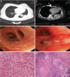
Oncocytic carcinoid tumor of the lung complicated by tuberculosis
- PMID: 31306231
- PMCID: PMC6759137
- DOI: 10.1097/CM9.0000000000000339
Oncocytic carcinoid tumor of the lung complicated by tuberculosis
Figures

Similar articles
-
Cytopathology of oncocytic carcinoid tumor of the lung mimicking granular cell tumor. A case report.Acta Cytol. 2000 Mar-Apr;44(2):247-50. doi: 10.1159/000326369. Acta Cytol. 2000. PMID: 10740615
-
[Oncocytic carcinoid tumor of the lung].Magy Seb. 2001 Feb;54(1):54-6. Magy Seb. 2001. PMID: 11299867 Hungarian.
-
Combined Typical Carcinoid-Adenocarcinoma Lung Tumor.J Bronchology Interv Pulmonol. 2017 Apr;24(2):e23-e25. doi: 10.1097/LBR.0000000000000359. J Bronchology Interv Pulmonol. 2017. PMID: 28323740 No abstract available.
-
Oncocytic carcinoid of the lung.Jpn J Thorac Cardiovasc Surg. 2005 Jul;53(7):393-6. doi: 10.1007/s11748-005-0058-y. Jpn J Thorac Cardiovasc Surg. 2005. PMID: 16095243 Review.
-
[Smoking and lung cancer].Duodecim. 1970;86(8):426-45. Duodecim. 1970. PMID: 4912052 Review. Finnish. No abstract available.
References
-
- Turan O, Ozdogan O, Gurel D, Onen A, Kargi A, Sevinc C. Growth of a solitary pulmonary nodule after 6 years diagnosed as oncocytic carcinoid tumour with a high 18-fluorodeoxyglucose (18F-FDG) uptake in positron emission tomography-computed tomography (PET-CT). Clin Respir 2013; 7:e1–e5. doi: 10.1111/j.1752-699X.2011.00274.x. - PubMed
-
- Varol Y, Varol U, Unlu M, Kayaalp I, Ayranci A, Dereli MS, et al. Primary lung cancer coexisting with active pulmonary tuberculosis. Int J Tuberc Lung Dis 2014; 18:1121–1125. doi: 10.5588/ijtld.14.0152. - PubMed
-
- Rihawi A, Huang G, Al-Hajj A, Bootwala Z. A case of tuberculosis and adenocarcinoma coexisting in the same lung lobe. Int J Mycobacteriol 2016; 5:80–82. doi: 10.1016/j.ijmyco.2015.07.001. - PubMed
-
- Morales-García C, Parra-Ruiz J, Sánchez-Martínez JA, Delgado-Martín AE, Amzouz-Amzouz A, Hernández-Quero J. Concomitant tuberculosis and lung cancer diagnosed by bronchoscopy. Int J Tuberc Lung Dis 2015; 19:1027–1032. doi: 10.5588 /ijtl d.14.0578. - PubMed
-
- Teng YL, Zou YB, Yan X, Cao DB, Guo L. Pulmonary oncocytic carcinoid-a case report. Chin J Pathol 2015; 44:344–345. doi: 10.3760/cma.j.issn. 0529-5807.2015.05.014. - PubMed
Publication types
MeSH terms
LinkOut - more resources
Full Text Sources
Medical

