Case Reports
. 2021 Nov 1;15(6):694-701.
doi: 10.1097/ICB.0000000000000891.

ENHANCED S-CONE SYNDROME: VISUAL FUNCTION, CROSS-SECTIONAL IMAGING, AND CELLULAR STRUCTURE WITH ADAPTIVE OPTICS OPHTHALMOSCOPY

Affiliations
Case Reports

ENHANCED S-CONE SYNDROME: VISUAL FUNCTION, CROSS-SECTIONAL IMAGING, AND CELLULAR STRUCTURE WITH ADAPTIVE OPTICS OPHTHALMOSCOPY

Michael J Ammar et al. Retin Cases Brief Rep. .

Abstract

Purpose: To describe in detail the phenotype of a patient with enhanced S-cone syndrome.

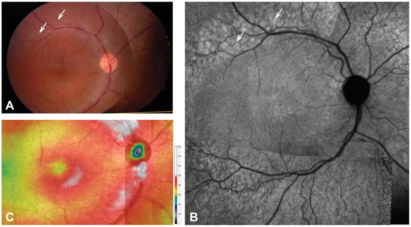
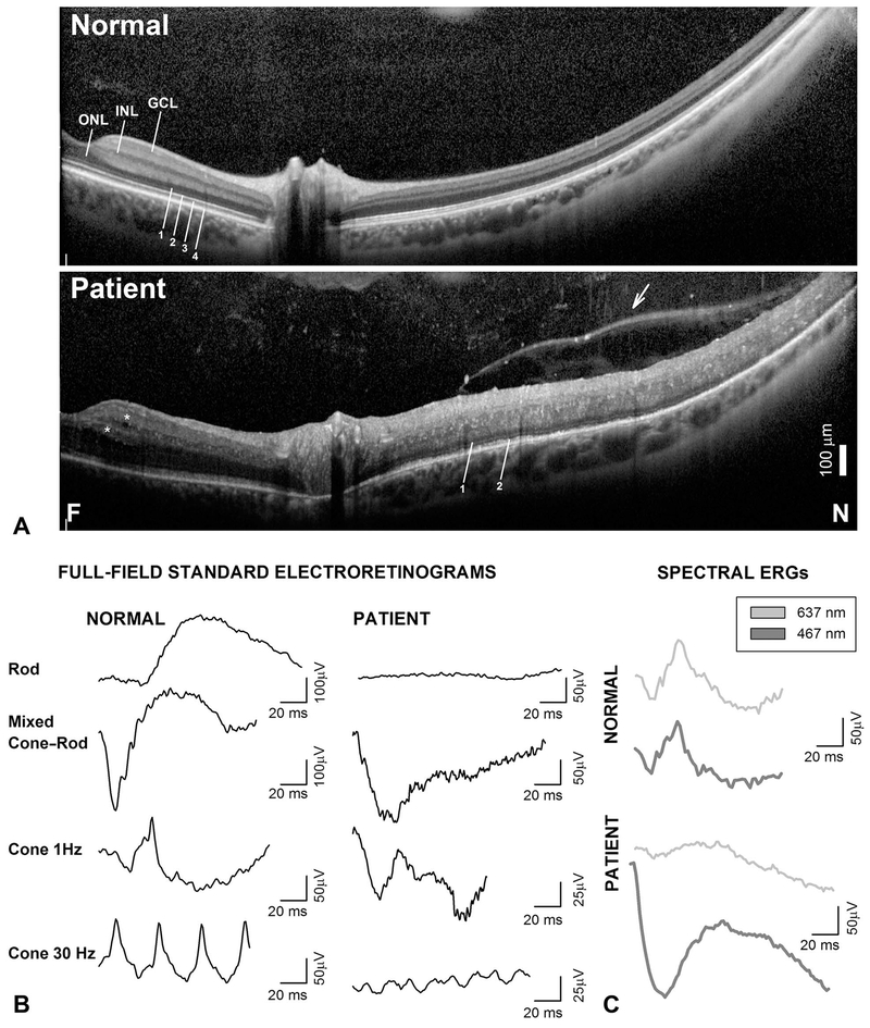
Methods: We describe a 13-year-old boy who presented with blurred vision, vitreous cells, cystoid macular edema refractory to steroid treatment, and a negative uveitic workup. The patient underwent a complete ophthalmic examination, full-field electroretinograms (ffERG), automatic static perimetry and multimodal imaging with spectral domain optical coherence tomography, and adaptive optics scanning laser ophthalmoscopy (AOSLO).

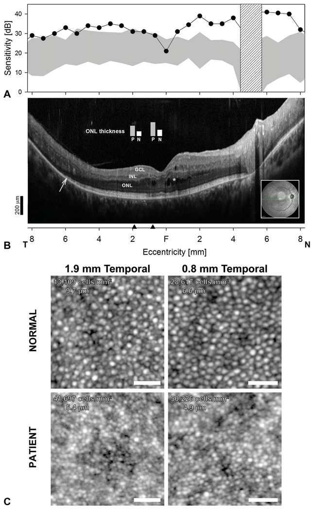
Results: Spectral domain optical coherence tomography demonstrated cystoid macular edema and a hyperthick, delaminated midperipheral retina. Fluorescein angiography did not demonstrate macular leakage. Rod-mediated ffERGs were undetectable, and there was a supernormal response to short-wavelength stimuli compared with photopically matched longer wavelengths of light consistent with enhanced S-cone syndrome. Gene screening was positive for compound heterozygous mutations NR2E3: a known (c.119-2 A>C) and a novel (c.119-1G>A) mutation. By perimetry, sensitivities were normal or above normal for short-wavelength stimuli; there was no detectable rod-mediated vision. AOSLO demonstrated higher than normal cone densities in the perifoveal retina and evidence for smaller outer segment cone diameters.

Conclusion: Evidence for supernumerary cones (at least twice the normal complement) by AOSLO and spectral domain optical coherence tomography was associated with supernormal S-cone sensitivities and electroretinogram responses confirming previous in vivo findings in postmortem human specimens. Smaller than normal cones in enhanced S-cone syndrome may represent "hybrid" photoreceptors analogous to the rd7/rd7 murine model of the disease.


Conflict of interest statement

J. I. W. Morgan is an inventor on US Patent 8226236. The remaining authors have any financial/conflicting interests to disclose.

Figures

Fig. 1.
Fig. 1.
En face imaging in the patient. Montages of fundus photographs (A), NIR-FAF (B), and total retinal thickness topography of the central macula and peripapillary retina (C). Arrows (A and B) point to the retina along the vascular arcades that appears normal on fundus photography but show hyperautofluorescence on NIR-FAF.
Fig. 2.
Fig. 2.
Diagnostic hallmarks of ESCS on SD-OCT and full-field ERGs. A. Horizontal, 16.4-mm-long, SD-OCT cross-section from near fovea (F) into nasal (N) retina in the patient compared with a normal subject. Nuclear layers (INL, inner nuclear layer; GCL, ganglion cell layer) and outer photoreceptor laminae are labeled (1, outer limiting membrane; 2, inner segment/outer segment junction near the inner segment ellipsoid zone [ISe]; 3, interdigitation between photoreceptor outer segment tips and apical retinal pigment epithelium; and 4, retinal pigment epithelium, following conventional terminology. Asterisks are placed near hyporreflective cystoid spaces. Arrow points to vitreoretinal band or posterior hyaloid that separates optically empty spaces from a vitreous with reflective dots. B. Standard full-field ERGs in the patient compared with a representative normal subject. C. Light-adapted ERGs elicited with long-wavelength (637-nm, thin gray traces) and SW (467-nm, thick gray traces) stimuli matched (3 phot-cd·s·m−2) for L-M cone stimulation in the normal subject elicit grossly mismatched waveforms in the patient. Traces start at stimulus onset. Calibration bars are to the right of the traces.
Fig. 3.
Fig. 3.
A. Central retinal function and colocalized cross-sectional and cellular structure in ESCS. S-cone sensitivity profiles measured with a 440-nm stimulus on a yellow L-M cone adapting background compared with the normal range (gray band = mean ± 2 SD). Hatched bar: blind spot. B. Horizontal, 16.5-mm, SD-OCT through the fovea in the patient. Inset is an NIR-REF image with the location and orientation of the scan (green arrow). Asterisks denote cystoid spaces. Arrow points to transition zone where the ISe band becomes undetectable. Black arrow heads on the eccentricity axis denote locations where ONL thickness measurements from the patient (P) were compared with normal measurements (N) (plotted as vertical bars overlaid above SD-OCT cross-section). Scale bar is to the bottom left; (A and B) share the same eccentricity scale. C. AOSLO images from the patient with ESCS compared with a normal. Cone densities and cone spacing are denoted in panel. Cone density is approximately 2- and 3-fold higher than normal in the patient at 0.8 mm and 1.9 mm (~3° and 6°) temporal to the fovea, respectively. Scale bar: 25 μm.

References

    1. Marmor M, Jacobson S. Diagnostic clinical findings of a new syndrome with night blindness, maculopathy, and enhanced S cone sensitivity. Am J Ophthalmol 1990;110:124–134. - PubMed
    1. Swaroop A, Xu JZ, Pawar H, et al. A conserved retina-specific gene encodes a basic motif/leucine zipper domain. Proc Natl Acad Sci U S A 1992;89:266–270. - PMC - PubMed
    1. Haider NB, Jacobson SG, Cideciyan AV, et al. Mutation of a nuclear receptor gene, NR2E3, causes enhanced S cone syndrome, a disorder of retinal cell fate. Nat Genet 2000;24:127–131. - PubMed
    1. Swaroop A, Kim D, Forrest D. Transcriptional regulation of photoreceptor development and homeostasis in the mammalian retina. Nat Rev Neurosci 2010;11:563–576. - PMC - PubMed
    1. Littink K, Stappers P, Riemslag F, et al. Autosomal recessive NRL mutations in patients with enhanced S-cone syndrome. Genes 2018;9:68. - PMC - PubMed

Publication types

MeSH terms

Supplementary concepts