Review
. 2020 Jan;40(1):38-48.
doi: 10.1002/pd.5526. Epub 2019 Jul 28.

Placental MRI and its application to fetal intervention

Affiliations
Review

Placental MRI and its application to fetal intervention

Rosalind Aughwane et al. Prenat Diagn. 2020 Jan.

Abstract

Objective: Magnetic resonance imaging (MRI) of placental invasion has been part of clinical practice for many years. The possibility of being better able to assess placental vascularization and function using MRI has multiple potential applications. This review summarises up-to-date research on placental function using different MRI modalities.

Method: We discuss how combinations of these MRI techniques have much to contribute to fetal conditions amenable for therapy such as singletons at high risk for fetal growth restriction (FGR) and monochorionic twin pregnancies for planning surgery and counselling for selective growth restriction and transfusion conditions.

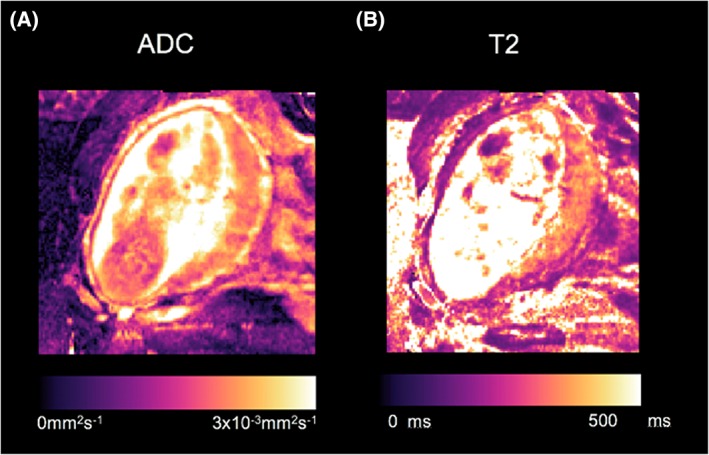
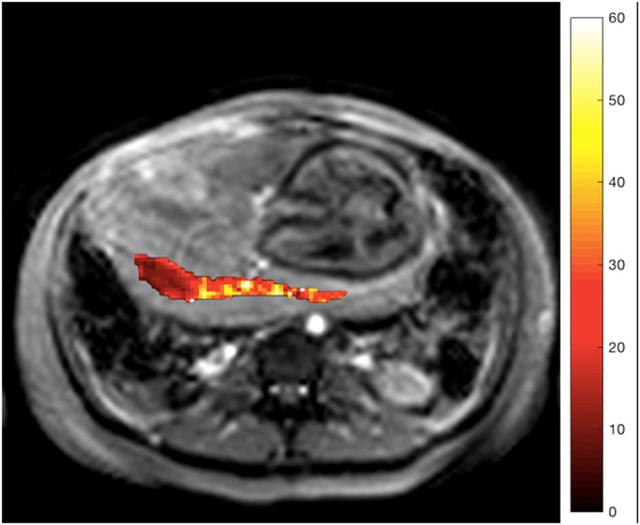
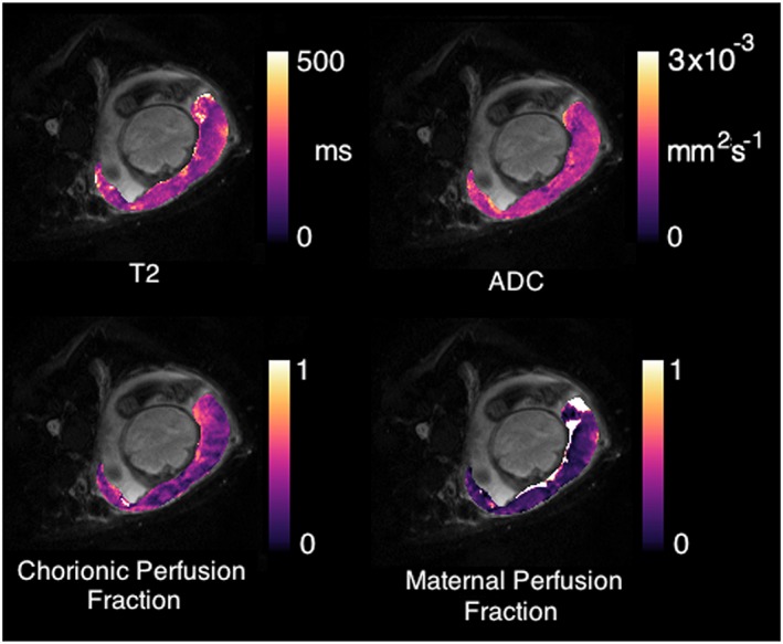
Results: The whole placenta can easily be visualized on MRI, with a clear boundary against the amniotic fluid, and a less clear placental-uterine boundary. Contrasts such as diffusion weighted imaging, relaxometry, blood oxygenation level dependent MRI and flow and metabolite measurement by dynamic contrast enhanced MRI, arterial spin labeling, or spectroscopic techniques are contributing to our wider understanding of placental function.

Conclusion: The future of placental MRI is exciting, with the increasing availability of multiple contrasts and new models that will boost the capability of MRI to measure oxygen saturation and placental exchange, enabling examination of placental function in complicated pregnancies.


Conflict of interest statement

We have no conflicts of interest to report.

Figures

Figure 1
Figure 1
Use of MRI in human placental conditions other than accreta, papers discussed in this review. Abbreviations in text [Colour figure can be viewed at http://wileyonlinelibrary.com]
Figure 2
Figure 2
MRI of placenta from a normally grown (left) and FGR (right) fetus. The placenta are marked with white stars. Note the difference in appearance in T2 weighted imaging, with the normal placenta appearing lighter in colour and more homogeneous
Figure 3
Figure 3
Example of placental single‐compartment ADC and T2 maps generated by linear least‐squares fitting [Colour figure can be viewed at http://wileyonlinelibrary.com]
Figure 4
Figure 4
T2 weighted structural image of axial slice through maternal abdomen, demonstrating uterine cavity, fetus, and placenta. Superimposed R2* map of the placental ROI (s−1) [Colour figure can be viewed at http://wileyonlinelibrary.com]
Figure 5
Figure 5
Physiological model‐fitting of the placenta.72 Parametric maps can be produced corresponding to fetal and maternal perfusion fractions (bottom row) simultaneously to conventional ADC and T2 maps (top row) [Colour figure can be viewed at http://wileyonlinelibrary.com]
Figure 6
Figure 6
Dynamic enhancement of the placenta with DCE‐MRI. Baseline image (1), arrival and wash‐in (2‐4), wash‐out (5‐6)

Similar articles

Cited by

References

    1. Crispi F, Miranda J, Gratacós E. Long‐term cardiovascular consequences of fetal growth restriction: biology, clinical implications, and opportunities for prevention of adult disease. Am J Obstet Gynecol. 2018;218(2):S869‐S879. 10.1016/j.ajog.2017.12.012 - DOI - PubMed
    1. Akolekar R, Bower S, Flack N, Bilardo CM, Nicolaides KH. Prediction of miscarriage and stillbirth at 11‐13 weeks and the contribution of chorionic villus sampling. Prenat Diagn. 2011;31(1):38‐45. 10.1002/pd.2644 - DOI - PubMed
    1. Akolekar R, Tokunaka M, Ortega N, Syngelaki A, Nicolaides KH. Prediction of stillbirth from maternal factors, fetal biometry and uterine artery Doppler at 19–24 weeks. Ultrasound Obstet Gynecol. 2016;48(5):624‐630. 10.1002/uog.17295 - DOI - PubMed
    1. Lawn JE, Blencowe H, Pattinson R, et al. Stillbirths: Where? When? Why? How to make the data count? Lancet. 2011;377(9775):1448‐1463. 10.1016/S0140-6736(10)62187-3 - DOI - PubMed
    1. Figueras F, Caradeux J, Crispi F, Eixarch E, Peguero A, Gratacos E. Diagnosis and surveillance of late‐onset fetal growth restriction. Am J Obstet Gynecol. 2018;218(2):S790‐S802.e1. 10.1016/j.ajog.2017.12.003 - DOI - PubMed

MeSH terms